
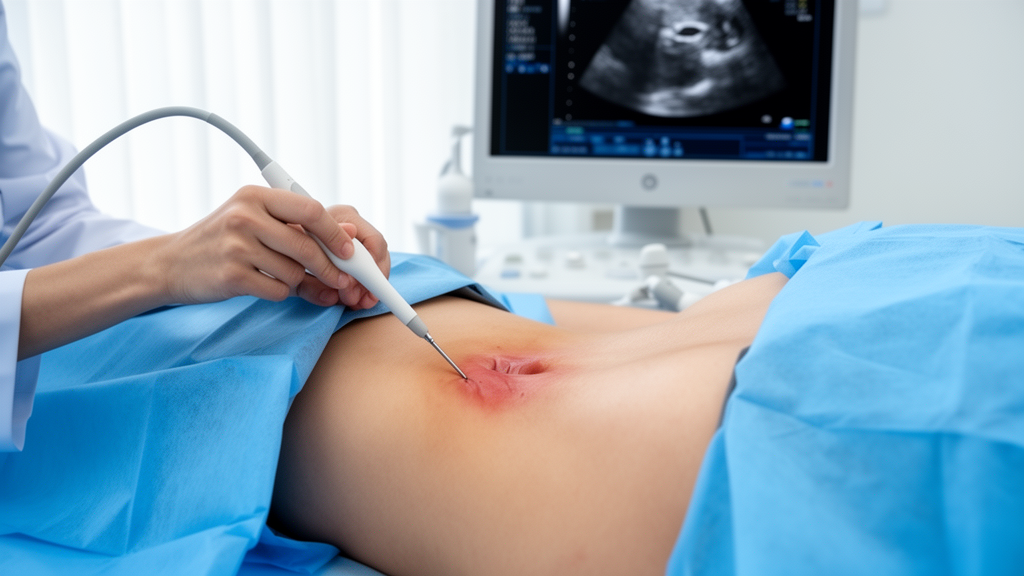
Ultrasound-guided sacroiliac joint puncture was associated with clinical improvement in 83.9% of 155 procedures performed on 130 patients with sacroiliac pain attributed to osteoarthritis or spondyloarthritis in a retrospective, single-centre study. The procedure-level improvement rate was 92.2% for the 77 procedures performed in patients with spondyloarthritis and 75.6% for the 78 procedures performed in patients with osteoarthritis, a statistically significant difference. Multivariable analysis identified diagnosis as the only independent predictor of improvement, with spondyloarthritis associated with 4.22 times the odds of improvement compared with osteoarthritis.
Researchers reviewed records for procedures carried out between June 2015 and June 2025 at a rheumatology centre in Madrid where the technique had been used routinely since 2012. Although the study was presented as an 11-year clinical experience, the analyzable cohort covered the decade for which electronic records provided systematic access to demographic, technical, clinical and safety information. Two rheumatologists with expertise in musculoskeletal ultrasound and ultrasound-guided interventions performed all punctures. Two researchers independently reviewed the records using a standardized data-collection protocol. Mean age was 48.7 years, with a standard deviation of 6.3 years and a range of 29 to 81 years.
Every patient underwent magnetic resonance imaging of the sacroiliac joints, which served as the principal diagnostic tool for attributing pain to the joint and distinguishing osteoarthritis from inflammation. Experienced musculoskeletal radiologists reviewed the images. Degenerative sacroiliac disease was characterized by findings such as joint-space narrowing, subchondral sclerosis, osteophytes and irregularity of the articular margins. Active sacroiliitis was defined according to ASAS criteria through bone-marrow edema on STIR sequences in typical locations. All patients classified as having spondyloarthritis fulfilled ASAS classification criteria for axial or peripheral disease. Two patients had both degenerative and inflammatory MRI changes; after clinical correlation and assessment against the classification criteria, both were assigned to the spondyloarthritis group.
The 155 procedures comprised 78 undertaken for sacroiliac osteoarthritis and 77 for spondyloarthritis. Within the inflammatory group, 63 procedures involved patients with axial spondyloarthritis, nine with psoriatic arthritis, three with inflammatory bowel disease-related enthesopathy and two with reactive arthritis. Thirty procedures were repeated interventions performed in 25 patients, while five were bilateral procedures conducted during the same session. Demographic variables were analysed per patient, whereas clinical outcomes were analysed per procedure. A complementary analysis restricted to the first procedure for each patient produced findings similar to those from the full procedure-level analysis, although the headline improvement rates of 92.2% and 75.6% were based on procedures.
All punctures used an upper, or cephalic, ultrasound-guided approach. With the patient lying face down on a rigid stretcher, the operator identified the fifth lumbar spinous process using a 6–9 MHz linear probe. The probe was displaced 1–2 cm caudally and 2–3 cm toward the side of the intended sacroiliac joint, then rotated slightly to accommodate the orientation of the joint’s upper and posterior aspects. After the target was visualized and the skin sterilized, a needle was advanced along the long axis of the probe, from medial to lateral at an inclination of 30–45 degrees until its bezel reached the joint region. The posterosuperior portion of the joint contains a relatively high proportion of ligamentous tissue, but it is also the most superficial segment and offers a useful acoustic window with limited needle travel. An approach toward the inferior pole instead places the trajectory near the sacral foramina and associated neurovascular structures.
Needle gauge and length were selected according to body habitus and the estimated distance between the skin and joint. Standard procedures used 21-gauge needles measuring 30–40 mm, whereas 20-gauge lumbar-puncture needles measuring 90 mm were used for patients with a high body mass index or when the estimated skin-to-joint distance exceeded 30–40 mm. The total injected volume did not exceed 1 mL. Triamcinolone, 20 mg in 0.5 mL, was used in all but three procedures in which it was unavailable; those cases received betamethasone, 5.7 mg in 0.5 mL. A local anesthetic could be added in cases of severe pain because the fibrous sacroiliac joint capsule is not highly permeable. No patient underwent puncture to collect joint fluid or for another diagnostic purpose, and no procedure involved an injected substance other than corticosteroid with or without anesthetic.
Clinical assessment combined the treating physician’s global impression of improvement with changes in pain intensity on a 0–10 visual analogue scale, or VAS. Follow-up assessment occurred at the first visit four to 12 weeks after the intervention. A procedure was classified as improved if the physician recorded a positive clinical judgment or the VAS score decreased. Stable symptoms were classified as no change, while procedures for which no outcome was documented were grouped with no change in the principal analysis. Clinical examination included standardized sacroiliac provocation tests: FABER, Gaenslen, thigh thrust, and compression and distraction. Clinicians also considered differential diagnoses involving facet-joint pain, discogenic pain and radicular pain. Each procedure was followed for as long as 12 months for safety observation, with events reported within 90 days classified as procedure-related for that analysis.
Clinical improvement was recorded for 130 of the 155 procedures, or 83.9%. Twenty-three procedures, representing 14.8%, were classified as having no change or no documented outcome, and two, or 1.3%, were associated with worsening. Pre- and post-procedure VAS scores were available for 58 procedures; across those cases, pain decreased by a mean 5.7 points with a standard deviation of 3.0. The mean interval between two procedures in the same patient was 110 days. In multivariable logistic regression, spondyloarthritis remained the only independent predictor of clinical improvement, with an adjusted odds ratio of 4.22, a 95% confidence interval of 1.22–14.58 and a p-value of 0.023. Obesity showed a nonsignificant trend toward lower improvement, while fibromyalgia showed a nonsignificant trend toward greater variability in outcomes; neither was independently associated with improvement. Age and sex were also not associated with the measured response.
No severe adverse event, bleeding event or hematoma was recorded, including among patients taking anticoagulants. One patient was admitted to hospital with suspected infection, but two sterile joint aspirations ruled out infectious arthritis. In six of the 155 procedures, or 3.9%, patients experienced enough immediate pain to require up to five minutes of observation. The investigators considered this immediate pain part of the expected course and found no association with short- or long-term outcomes. All patients said they would undergo the procedure again if necessary. Patients taking acenocoumarol underwent INR testing and received injections only when the value was 3.0 or lower, without interrupting anticoagulation. Dabigatran dosing was managed according to renal function: treatment was withheld for 24 hours before the procedure and restarted the following day. No procedure was suspended or delayed because of pain or another cause, and each took 10–15 minutes.
These results support the technical feasibility of ultrasound-guided sacroiliac puncture and show an observed safety profile that included no severe complications in the cohort. They do not establish that the injection was more effective than placebo, blind puncture or another treatment. The retrospective design, absence of a control group and reliance on clinical records leave room for placebo effects, regression to the mean, the natural history of sacroiliac pain and changes in concurrent treatment. The physician-based outcome measure may also have included modest changes that patients did not regard as clinically meaningful, while the lack of extended VAS follow-up prevented the investigators from determining how long pain relief lasted. Ultrasound improves needle visualization compared with landmark-only puncture, but it cannot confirm intra-articular spread as directly as fluoroscopy or computed tomography, so injectate may have reached periarticular or ligamentous tissue rather than the joint space. Retrospective retrieval from records may also have underestimated mild or systemic corticosteroid effects. The results therefore show an association between diagnosis and clinical improvement rather than proof that the procedure changes disease progression or delays escalation of therapy.
Subject of Research: Clinical outcomes and safety of ultrasound-guided sacroiliac joint corticosteroid puncture for osteoarthritis and spondyloarthritis
Article Title: Feasibility of ultrasound-guided sacroiliac joint puncture in the treatment of inflammatory and osteoarthritis pain: a 11-years single-centre experience
Article References: Guillén-Astete, C. A., Kanaffo, S., Andreu-Suarez, Á., Urrego-Laurín, C., Blanco-Cáceres, B., & Zurita-Prada, P. A. (2026). Feasibility of ultrasound-guided sacroiliac joint puncture in the treatment of inflammatory and osteoarthritis pain: a 11-years single-centre experience. Frontiers in Medicine, 13, Article 1997507. https://doi.org/10.3389/fmed.2026.1997507
Image Credits: AI Generated
DOI: 10.3389/fmed.2026.1997507
Keywords: ultrasound-guided puncture, sacroiliac joint, spondyloarthritis, osteoarthritis, corticosteroid injection, clinical improvement, retrospective study, treatment safety, pain outcomes, musculoskeletal ultrasound, Feasibility, ultrasound-guided