
A prospective clinical study has established that single-voxel edited magnetic resonance spectroscopy conducted on a clinical 3T scanner can reliably characterize key redox metabolites noninvasively in patients diagnosed with isocitrate dehydrogenase-wildtype glioblastoma. Isocitrate dehydrogenase-wildtype glioblastoma represents the most frequent and aggressive primary central nervous system malignancy in adults, consistently linked to poor clinical prognosis and limited therapeutic responsiveness. Within the challenging tumor microenvironment, neoplastic cells depend heavily on cellular antioxidant machinery to buffer elevated levels of oxidative stress and sustain uncontrolled cellular proliferation. Central to this defense mechanism is glutathione, the principal intracellular antioxidant, alongside its upstream biosynthetic precursor cystathionine. The newly reported findings demonstrate that specialized spectral editing can resolve these compounds in vivo, providing quantitative insights into how aggressive brain tumors orchestrate their internal antioxidant reserves.
The biological rationale for investigating metabolic redox pathways in isocitrate dehydrogenase-wildtype glioblastoma stems from the central role that reactive oxygen species play in tumor biology. In this lethal glioma entity, distinct somatic mutations, chronic local inflammation, and elevated immune signaling elevate oxidative pressure throughout neoplastic tissue. Cytokines such as tumor necrosis factor alpha stimulate reactive oxygen species generation, which paradoxically triggers redox-sensitive signaling cascades, including the PI3K and Akt pathways, thereby facilitating cytoskeletal alterations, cellular migration, survival, and invasive growth. To avoid catastrophic self-destruction from excessive oxidative stress, neoplastic cells rely on glutathione, a tripeptide synthesized from glutamate, cysteine, and glycine. The sulfhydryl moiety of cysteine provides the essential reducing capability required for neutralizing free radicals. Because intracellular cysteine supply frequently limits glutathione production, tumor cells upregulate the transsulfuration pathway, wherein cystathionine is converted into cysteine by the enzyme cystathionine-gamma-lyase. Consequently, evaluating both cystathionine and glutathione provides a direct window into tumor metabolic flexibility.
To investigate these metabolic dynamics in living human subjects, the research team designed a prospective study conducted at a tertiary care medical center between January 2023 and July 2025. The study protocol was approved by the local institutional review board under project number 2022-909 in adherence to the Helsinki Declaration, with written informed consent obtained from every participant. The trial was formally entered into the German Clinical Trials Register under registration identifier DRKS00032097. The investigation was structured in two distinct phases: an initial validation phase assessing scan-rescan reproducibility in healthy volunteers, followed by the evaluation of adult patients presenting with magnetic resonance imaging evidence of suspected diffuse glioma who were scheduled to undergo neurosurgical biopsy or tumor resection.
Rigorous diagnostic and methodological screening criteria were applied to construct a uniform clinical cohort for final spectral evaluation. Among the 27 enrolled patients with suspected gliomas, 4 individuals were excluded after histological and methylation analyses failed to confirm adult-type diffuse glioma; definitive diagnoses revealed non-small cell lung cancer metastasis, localized cerebral inflammation, dysembryoplastic neuroepithelial tumor, and a canceled surgical indication. This left 18 patients confirmed with World Health Organization central nervous system grade 4 isocitrate dehydrogenase-wildtype glioblastoma. Following spectroscopic data collection, an additional 3 cases were excluded from final analysis due to unacceptable spectral degradation, including spurious echo contamination, severe subtraction artifacts, and excessive metabolite linewidth exceeding 0.1 parts per million, leaving a final evaluable glioblastoma cohort of 15 patients.
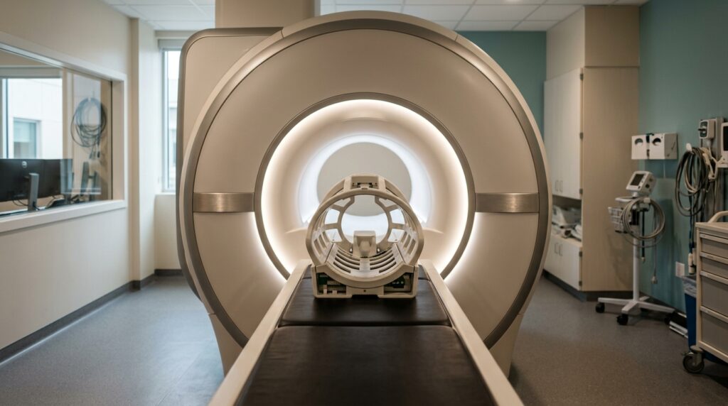
All magnetic resonance examinations were carried out on a 3T scanner equipped with a 20-channel head receiver coil, employing a specialized Mescher-Garwood semi-adiabatic localization by adiabatic selective refocusing sequence known as MEGA-sLASER. To make the procedure clinically viable, spectroscopic scans maintained a short acquisition duration of approximately 6.44 minutes, accumulating 192 total transients across three interleaved editing states. Voxel sizes ranged between 8 and 16 cubic centimeters to minimize partial-volume contamination while preserving adequate signal-to-noise ratios. The interleaved sequence applied frequency-selective radiofrequency editing pulses at 1.9 parts per million to simultaneously target gamma-aminobutyric acid and cystathionine, at 4.56 parts per million to selectively edit glutathione resonances, and at 7.5 parts per million as an unedited reference condition. An unsuppressed water reference spectrum was collected using 4 transients for quantification, with repetition and echo times set at 2000 and 80 milliseconds, respectively.
Prior to clinical patient measurements, the reproducibility of the MEGA-sLASER protocol was rigorously demonstrated in 5 healthy volunteers who completed two consecutive scanning sessions separated by a 15-minute recess outside the scanner bore. Because cystathionine concentrations are negligibly low in healthy brain parenchyma, gamma-aminobutyric acid served as the primary co-edited marker for repeatability. Analysis demonstrated consistent signal-to-noise ratios, narrow linewidths, and stable spectral fitting uncertainties between sessions. Gray matter voxel fractions did not significantly differ between repetitions, averaging 0.39 plus or minus 0.11 in the first scan and 0.38 plus or minus 0.09 in the second. Paired comparisons revealed no statistically significant differences in quantified metabolite concentrations: total gamma-aminobutyric acid measured 3.60 plus or minus 0.73 millimolar in session one versus 3.33 plus or minus 0.31 millimolar in session two, while glutathione concentrations were 0.37 plus or minus 0.27 millimolar compared to 0.49 plus or minus 0.18 millimolar. Bland-Altman analyses confirmed narrow limits of agreement, yielding repeatability coefficients of 1.26 millimolar for gamma-aminobutyric acid and 0.53 millimolar for glutathione.
When applied to glioblastoma patients, spectral acquisitions targeted both contrast-enhancing neoplastic tissue and corresponding normal-appearing parenchyma in the contralateral cerebral hemisphere. Tissue segmentation revealed that tumor voxels contained an average fraction of 0.74 plus or minus 0.22 composed of viable tumor tissue and peritumoral subregions, with necrotic elements accounting for 0.07 plus or minus 0.11. In contralateral control voxels, healthy gray matter and white matter fractions averaged 0.30 plus or minus 0.10 and 0.62 plus or minus 0.13, respectively. Spectral quality measures derived from unedited spectra remained closely matched across regions, showing linewidths of 0.05 plus or minus 0.02 parts per million in tumor tissue and 0.06 plus or minus 0.02 parts per million in contralateral tissue, with respective signal-to-noise ratios of 14.5 plus or minus 5.32 and 16.1 plus or minus 5.15.
Metabolite comparisons using linear mixed models revealed marked biochemical shifts between tumoral and normal brain tissue. Neoplastic tissue exhibited a significant elevation in cystathionine levels, reaching 1.17 plus or minus 1.30 millimolar compared to 0.63 plus or minus 0.59 millimolar in contralateral voxels, representing a statistically significant increase with a p-value of 0.03. In contrast, total gamma-aminobutyric acid was significantly depleted within tumor tissue, measuring 2.36 plus or minus 0.70 millimolar compared to 3.04 plus or minus 0.89 millimolar in contralateral tissue, reflecting a highly significant difference with a p-value of 0.006. Glutathione concentrations averaged 0.61 plus or minus 0.54 millimolar in tumors versus 0.38 plus or minus 0.25 millimolar in contralateral brain, which did not attain statistical significance as a uniform group difference due to high intertumoral variability across the glioblastoma patient cohort.
To unravel the substantial individual variability observed in tumoral glutathione, the investigators conducted exploratory multiple linear regression incorporating anatomic subregion volumes and co-quantified metabolites. Automated volumetric segmentation classified tumors into enhancing tumor, non-enhancing tumor core, and surrounding non-enhancing FLAIR hyperintensity. The resulting linear regression model explained 96 percent of the variance in tumoral glutathione concentration, yielding an adjusted R-squared of 0.96. Glutathione concentration demonstrated strong, statistically significant positive associations with enhancing tumor fraction, showing a regression coefficient beta of 0.97 with standard error 0.17 and 95 percent confidence interval from 0.59 to 1.36, total choline concentration with beta of 0.13, and cystathionine concentration with beta of 0.26. Conversely, non-enhancing tumor core volume and total N-acetyl aspartate exhibited no statistically significant associations with glutathione levels, confirming that antioxidant accumulation is heavily concentrated within cellular, metabolically active enhancing tumor zones.
The study further uncovered notable associations between spectroscopic redox measurements, immunohistochemical markers, and molecular tumor subtypes. Analysis of tumor protein p53 immunoreactivity revealed a significant negative correlation with intratumoral glutathione concentration, characterized by a Spearman rank correlation coefficient rho of minus 0.7 and a p-value of 0.008. Glutathione concentrations showed no significant differences when stratifying the 14 molecularly classified tumors by O6-methylguanine-DNA methyltransferase promoter methylation status, which returned a p-value of 0.66. However, significant variations emerged when comparing DNA methylation-based subclasses: tumors categorized as mesenchymal exhibited substantially lower glutathione concentrations, averaging 0.14 plus or minus 0.15 millimolar, whereas tumors classified as receptor tyrosine kinase I or II subtypes maintained markedly higher concentrations, averaging 0.65 plus or minus 0.49 millimolar, a distinction confirmed by a Wilcoxon rank-sum test p-value of 0.04.
The biological interpretations offered by the authors contextualize these metabolic shifts within broader glioma pathophysiology. The elevation of cystathionine aligns with upregulation of the transsulfuration pathway, which channels precursors into glutathione synthesis and bolsters cellular defense against oxidative injury induced by radiotherapy and alkylating chemotherapy. Regarding p53 dynamics, wild-type p53 normally maintains redox balance by modulating glutathione recycling and regulating the SLC7A11 subunit of the cystine antiporter system. Although early TP53 mutations lower basal glutathione and elevate mutagenic stress, established tumors often activate alternative pathways to restore antioxidant defense, while non-neoplastic stromal elements and cellular hypoxia can stabilize wild-type p53 protein, complicating immunohistochemical interpretations.
Despite these compelling results, the investigators highlighted technical challenges and clinical boundaries inherent to their protocol. Multiple alignment algorithms, including RobustSpecReg and SpecRegHERMES, had to be tailored individually to avoid subtraction artifacts. Most importantly, the modest final sample size of 15 glioblastoma patients limits statistical power, precluding definitive conclusions regarding long-term clinical survival or treatment response. The authors emphasize that larger multicenter prospective cohorts will be essential to validate these findings and formally assess whether in vivo edited MR spectroscopy can serve as a dependable imaging biomarker for tailoring redox-modulating brain tumor therapies.
Subject of Research: In vivo magnetic resonance spectroscopy characterization of redox metabolites in IDH-wildtype glioblastoma
Article Title: In vivo characterization of the redox balance in IDH-wildtype glioblastomas: a J-difference edited MEGA-sLASER MRS study at 3T
Article References: Alcicek, S., Manzhurtsev, A., Thomas, D. C., Divé, I., Weber, K. J., Prinz, V., Jussen, D., Deelchand, D. K., Oeltzschner, G., Ronellenfitsch, M. W., Steinbach, J. P., Hattingen, E., Pilatus, U., & Wenger, K. J. (2026). In vivo characterization of the redox balance in IDH-wildtype glioblastomas: a J-difference edited MEGA-sLASER MRS study at 3T. Cancer & Metabolism, 14(1), Article 10. https://doi.org/10.1186/s40170-026-00432-7
Image Credits: AI Generated
DOI: 10.1186/s40170-026-00432-7
Keywords: glioblastoma, magnetic resonance spectroscopy, glutathione, cystathionine, redox balance, neuro-oncology, molecular imaging, metabolism, vivo, characterization, redox, balance