
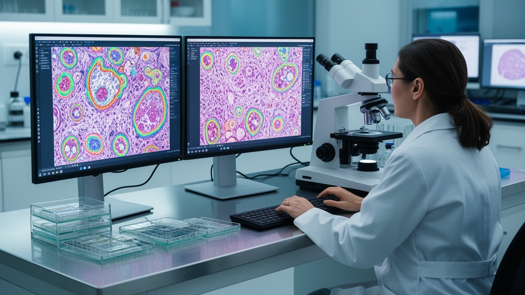
In an extensive evaluation of computational pathology techniques, deep learning algorithms operating without task-specific parameter fine-tuning demonstrated superior performance over classical thresholding and interactive pixel classification methods when segmenting cell nuclei across multiple cancer types. The findings emerge from a comprehensive narrative review and experimental benchmark published in Discover Artificial Intelligence by researchers examining computer vision applications in the four most prevalent solid malignancies worldwide: lung, breast, colorectal, and prostate cancers. Traditionally, the clinical gold standard for oncological verification has depended upon manual microscopic evaluation of tissue sections stained with hematoxylin and eosin, a routine process processing up to three million slides globally each day. However, standard visual assessment by human pathologists remains constrained by significant intratumoral heterogeneity, diagnostic subjectivity between observers, and high cognitive fatigue in high-volume pathology settings. To address these clinical challenges, modern computational frameworks seek to automate feature extraction, standardize morphological evaluations, and map complex spatial architectures within digitized whole-slide images.
The experimental portion of the research established a standardized five-tool comparative benchmark across five distinct dataset groups spanning four tumor types, utilizing image sets from the MoNuSAC, CoNSeP, and TNBC repositories. The authors specifically compared three distinct architectural paradigms running exclusively under default, non-fine-tuned configurations: deep learning models represented by Cellpose and the Fiji StarDist plugin, classical global thresholding implemented via Otsu thresholding in Icy and CellProfiler, and interactive machine learning represented by Ilastik running on a minimal annotation protocol of ten nuclei across three images. Across five of the six evaluated dataset cohorts, Cellpose achieved the highest segmentation accuracy, yielding superior Dice scores with ninety-five percent confidence intervals that did not overlap with StarDist. StarDist demonstrated slightly superior accuracy only on the sparsely cellular triple-negative breast cancer dataset, achieving a Dice coefficient of 0.84 compared to 0.82 for Cellpose.
The benchmark revealed distinct behavioral and architectural failure modes separating classical algorithms from deep learning frameworks. Tools relying on automated global thresholding, specifically Icy and CellProfiler, suffered from pronounced small-scale oversegmentation, producing elevated false-positive rates and depressed precision values ranging from 0.35 to 0.53 despite registering high recall scores between 0.75 and 0.85. The authors noted that this oversegmentation stemmed directly from the absence of explicit structural shape models, causing thresholding routines to fracture dense cell clusters and misclassify non-nuclear intercellular structures as nuclear material, particularly within highly cellular colorectal and lung tissue sections. Ilastik occupied an intermediate position, demonstrating greater stability in high-density tissue regions than unguided thresholding but frequently merging adjacent cell nuclei into large, contiguous conglomerates and mistakenly classifying stromal fragments as tumor nuclei in small-cell regions. Conversely, Cellpose combined high precision between 0.74 and 0.92 with competitive recall by incorporating deep morphological representations of cell contours, while StarDist exhibited more conservative segmentation boundaries that led to the omission of nuclei within tightly packed tissue areas.
Beyond nuclei segmentation, the authors emphasized that the primary biological stumbling block in digital oncology is the rigorous quantification of tumor heterogeneity, which manifests across diverse genetic, epigenetic, and spatial scales within a single neoplastic lesion. While next-generation sequencing provides deep genomic readouts, bulk molecular assays homogenize tissue samples and obliterate spatial distribution, whereas gross radiological modalities such as computed tomography and magnetic resonance imaging lack the microscopic resolution necessary to resolve subclonal microenvironments. Digitized histopathology preserves the intact geographic architecture of the tumor microenvironment, enabling computational systems to evaluate cellular atypia, stromal reactions, and immune infiltration simultaneously. The review structured heterogeneity quantification into a multi-scale analytical hierarchy, starting with cellular morphological indices such as nuclear pleomorphism descriptors and Haralick texture features derived from gray-level co-occurrence matrices. Moving upward in scale, spatial statistics like Moran’s I, Delaunay triangulation edge variances, and fractal dimensions quantify the disorder of tissue boundaries and the infiltrative complexity of the tumor-stroma interface, while patch-level clustering using Shannon diversity metrics and multi-scale dimensionality reduction construct comprehensive signatures of whole-slide architectural variance.
Tracing the technological trajectory of digital pathology, the review detailed the ongoing shift from early hand-engineered visual descriptors toward deep self-supervised networks and universal foundation models. Hand-crafted features based on color decomposition, such as isolating the hue channel in hue-saturation-value color space for hyperchromatic prostate nuclei or utilizing CIELAB chromogen channels for breast cancer immunohistochemistry scoring, provided initial objective baselines but struggled to capture complex, high-dimensional phenotypic phenotypes. Modern deep learning architectures, such as convolutional networks and vision transformers, automate this feature extraction process, though vision transformers typically require vast annotated corpora that are rarely accessible in public archives due to proprietary limitations and labor-intensive annotation requirements. To overcome these data bottlenecks, self-supervised learning algorithms train feature encoders on unlabeled whole-slide fragments using pretext tasks, while generative artificial intelligence architectures, including generative adversarial networks and diffusion models, are being explored to synthesize underrepresented histopathological patterns, such as rare micropapillary lung carcinoma or high-grade cribriform prostate lesions, and to harmonize staining variations across clinical centers.
This foundational evolution has culminated in the emergence of massive pathology foundation models trained on millions of tissue patches across diverse organ systems. Notable vision-only models highlighted in the review include UNI, which scaled from an initial 300-million parameter vision transformer to a 681-million parameter model trained across more than one hundred million patches, Virchow and Virchow 2, trained on up to 3.1 million whole-slide images spanning seventeen tissue types, and slide-level architectures like Prov-GigaPath. Simultaneously, vision-language models such as CONCH, pretrained on 1.17 million paired histopathology images and captions, and slide-level architectures like TITAN and mSTAR are aligning whole-slide visual features with clinical diagnostic text and genomic profiles. Independent benchmarking surveyed in the review demonstrated that vision-language architectures can match or exceed the predictive capabilities of vision-only models in biomarker prediction and morphological subtyping, providing a flexible framework where a single frozen foundational visual encoder can be adapted through task-specific aggregators to execute diverse diagnostic operations without requiring de novo model development.
To transition computational models from retrospective research into reliable clinical decision-support tools, the review presented a structured taxonomy aligning distinct computational vision tasks with the divergent diagnostic imperatives of the four solid tumor types. In lung adenocarcinoma, where diagnostic workflow focuses on identifying non-small cell subtypes and actionable driver mutations such as EGFR and ALK, slide-level multiple-instance learning frameworks serve to aggregate patch representations and prioritize diagnostic areas over surrounding necrotic tissue. Breast cancer assessment demands a hybrid framework integrating semantic segmentation of tissue architecture with high-precision object detection for mitotic counting and quantitative scoring of hormone receptor and HER2 expression. Colorectal cancer diagnostics center on microenvironmental interactions, requiring semantic segmentation of the invasive front combined with graph neural networks to map spatial topologies, such as lymphocyte proximity to tumor glands and tumor-stroma ratios. Conversely, prostate cancer management presents an extreme needle-in-a-haystack detection problem, requiring attention-weighted models tuned to locate sparse malignant foci and classify complex cribriform glandular patterns amidst extensive benign background architecture.
Because high-stakes clinical translation requires verifiable decision-making, the authors stressed that explainable artificial intelligence is an ethical and clinical prerequisite rather than an optional technical enhancement. Common post-hoc interpretability methods include gradient-weighted class activation mapping, which generates visual heatmaps highlighting influential whole-slide regions, and multi-head attention map visualizations within vision transformers that elucidate how models contextualize individual glands relative to surrounding tissue. Graph neural networks offer inherent structural transparency because their decision pathways can be traced along biological nodes and relational edges representing interacting cells and tissue compartments. The authors cautioned that model explainability must not rely solely on subjective visual plausibility; instead, explanations must undergo rigorous biological validation. This validation requires evaluating localization accuracy against expert annotations, measuring spatial overlap with verified histological structures, conducting failure-case analyses, and executing reader studies to confirm whether algorithmic explanations genuinely enhance pathologist diagnostic confidence and inter-observer agreement in accordance with formal reporting frameworks like the DECIDE-AI guidelines.
The review explicitly detailed several methodological and structural limitations that must temper the interpretation of current computational histopathology literature. Most published models, including foundation models and generative frameworks, remain restricted to retrospective evaluations on curated cohorts, with few prospective multi-center trials having demonstrated concrete improvements in patient outcomes. Furthermore, the primary literature is heavily vulnerable to selection bias because negative findings are rarely reported, no formal risk-of-bias assessment tools were applied across the narrative literature search, and review protocols were not prospectively registered. The authors also noted that their own comparative benchmark was intentionally restricted to out-of-the-box software performance using default parameters, excluding foundation models and vision-language systems because their deployment demands specialized programming pipelines, significant graphics processing cluster infrastructure, and lacks the accessible graphical interfaces required for routine clinical workstations. Additionally, generative models carry non-trivial clinical risks, including mode collapse during training and the synthesis of biologically implausible nuclear artifacts that could compromise diagnostic safety if used carelessly for data augmentation.
Achieving equitable, robust clinical deployment will ultimately depend on establishing multi-institutional validation cohorts, standardizing color normalization frameworks to overcome scanner variations, and designing human-in-the-loop systems that augment human pathologists rather than attempting autonomous diagnosis. Interactive platforms integrating large language models through retrieval-augmented generation and structured clinical templates could soon allow pathologists to query multimodal slide representations, automate standard reporting metrics, and uncover novel prognostic morphometric features across historical cohorts. However, the researchers concluded that computational histopathology can only realize its transformative potential in personalized oncology if future algorithms remain firmly grounded in biomedical realities, scale-agnostic tissue hierarchies, and transparent, clinically validated workflows designed to safeguard patient care.
Subject of Research: Computer vision and deep learning algorithms in histopathological cancer diagnostics
Article Title: Review of computer vision algorithms in histopathology to decipher complex features in cancer
Article References: Illarionova, S., Studenikina, A., Carreras, J., Zapevalina, M., Bernstein, A. V., Hamoudi, R., & Sharaev, M. (2026). Review of computer vision algorithms in histopathology to decipher complex features in cancer. Discover Artificial Intelligence, 6(1), Article 1752. https://doi.org/10.1007/s44163-026-02592-3
Image Credits: AI Generated
DOI: 10.1007/s44163-026-02592-3
Keywords: histopathology, computational pathology, computer vision, deep learning, whole slide imaging, cell nuclei segmentation, tumor heterogeneity, Cellpose, StarDist, hematoxylin and eosin, digital pathology, vision transformers, foundation models, explainable artificial intelligence, colorectal cancer, prostate cancer, breast cancer, lung adenocarcinoma
