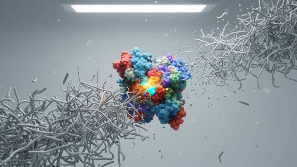
A newly characterized bacterial enzyme designated as Kgh3 effectively prevents and dismantles persistent Klebsiella pneumoniae biofilms through the direct enzymatic cleavage of cell wall peptidoglycan. In a laboratory study evaluating microbial control mechanisms, researchers discovered that Kgh3 functions as an active endolysin capable of inducing rapid bacterial lysis rather than merely targeting structural matrix polysaccharides. Investigations conducted across both reference strains and recalcitrant clinical isolates revealed that this glycoside hydrolase family 19 enzyme exerts robust dual-action capabilities, arresting nascent biofilm growth at early developmental stages and dismantling mature, established microbial communities. Crucially, the biological intervention demonstrated significant in vivo efficacy within an experimental mouse model of chronic dorsal wound infection, where administration of the purified enzyme accelerated tissue repair, prevented systemic weight loss, and substantially cleared localized bacterial colonization without generating cytotoxic injury in mammalian cell lines.
Klebsiella pneumoniae represents an opportunistic Gram-negative pathogen recognized by the World Health Organization as a critical-priority target for antimicrobial research due to its extensive multidrug resistance and propensity to colonize abiotic surfaces and host tissues. Pathogenic persistence in clinical environments frequently depends on the bacterium’s capacity to aggregate into dense, multicellular biofilms encased in a protective extracellular polymeric substance comprised primarily of polysaccharides, proteins, and extracellular DNA. This architectural matrix acts as a formidable physical and physiological shield, impeding immune surveillance and blunting the therapeutic impact of conventional antibiotic regimens. While conventional physical and chemical strategies such as photodynamic therapy or non-specific peptide applications can destabilize microbial conglomerates, they frequently produce non-target damage against healthy mammalian tissues and commensal microflora. Consequently, research initiatives have increasingly centered on identifying specialized catalytic proteins capable of degrading distinct structural constituents to disarm protected bacterial populations.
In the present study, investigators targeted Kgh3, an enzyme derived directly from the genome of the antimicrobial-resistant reference strain Klebsiella pneumoniae MGH78578. The coding sequence, cataloged under GenBank accession WP_012967893.1, was cloned into a pET-28a vector to generate an N-terminal hexahistidine-tagged fusion construct, which was subsequently expressed heterologously in Escherichia coli BL21 (DE3) cells under 0.5 mM isopropyl β-d-1-thiogalactopyranoside induction at 16 degrees Celsius. Purification using nickel-nitrilotriacetic acid affinity chromatography produced a yield of approximately 40 milligrams of homogeneous soluble protein per liter of culture. Following isolation, microtiter dish assays paired with crystal violet spectrophotometric staining were implemented to assess the enzyme’s capacity to modify biofilm dynamics. Quantitative dose-response analyses established that Kgh3 disrupted pre-existing biofilms of strain MGH78578 in a concentration-dependent manner, exhibiting a median effective concentration of 19.15 ± 2.47 micrograms per milliliter, while preventing initial biofilm establishment with a median effective concentration of 40.67 ± 6.16 micrograms per milliliter.
Enzymatic specificity was confirmed by engineering inactive site-directed variants designated E49A and E58A, both of which completely lost the capacity to dismantle mature biofilm matrices. In contrast, treatment of established MGH78578 biofilms with 100 micrograms per milliliter of native Kgh3 for two hours achieved a reduction in remaining biomass exceeding 95 percent. To explore whether catalytic performance required outer membrane permeabilizers, a common necessity for Gram-negative targeting endolysins, the investigators co-administered Kgh3 alongside ethylenediaminetetraacetic acid, Triton X-100, or chloroform. Subsequent thermal stability profiling indicated that Kgh3 retained 81.93 percent of its disruptive activity following one hour of incubation at 37 degrees Celsius, and preserved 55.06 percent residual activity after ten hours, highlighting an operational durability favorable for sustained downstream therapeutic applications.
To confirm that these enzymatic attributes were not restricted to a single domesticated laboratory isolate, the investigators screened 25 clinical isolates of Klebsiella pneumoniae and identified six robust biofilm-forming strains for extended testing. When exposed to 100 micrograms per milliliter of purified Kgh3, these clinical isolates exhibited significant reductions in both biofilm development and mature community persistence. Across the panel, Kgh3 disrupted established clinical biofilms with clearing efficiencies reaching up to 62.95 percent, while achieving preventative inhibition rates as high as 86.66 percent. Prior to examining therapeutic translation in whole organisms, the researchers verified the biocompatibility of the enzyme by performing comprehensive in vitro cytotoxicity screenings across multiple mammalian cell lineages. Cultured human embryonic lung fibroblasts, CCC-ESF-1 human embryonic skin fibroblasts, RAW 264.7 murine macrophages, and THP-1 human acute monocytic leukemia cells were incubated with enzyme concentrations escalating up to 500 micrograms per milliliter for 24 hours. Cell Counting Kit-8 colorimetric viability readouts showed only negligible fluctuations relative to untreated controls, confirming that Kgh3 lacks intrinsic cytotoxicity toward host cells.
Therapeutic utility was subsequently established using an in vivo dorsal wound infection model conducted in six- to eight-week-old male ICR mice. Surgical skin excisions measuring one centimeter in diameter were created under isoflurane anesthesia and local lidocaine analgesia, followed by subcutaneous inoculation with ten million colony-forming units of Klebsiella pneumoniae MGH78578. After six hours of infection to facilitate localized biofilm establishment, animals received therapeutic subcutaneous injections of 100 microliters of Kgh3 solution or sterile phosphate-buffered saline, with daily dressing changes and identical repeated administrations throughout a seven-day observation course. Infected control mice treated exclusively with vehicle experienced rapid weight loss ranging between 10 and 13 percent within 24 hours and sustained poor healing indicative of persistent chronic infection. Conversely, animals receiving Kgh3 therapy exhibited only a transient five percent weight reduction on day one, comparable to uninfected controls, and exhibited steady weight recovery alongside visibly accelerated wound closure across the monitoring window.
Microbiological and histological evaluations of wound beds harvested on day seven post-infection confirmed substantial clinical improvement in enzyme-treated mice. Quantitative serial-dilution colony counts performed on Luria-Bertani agar demonstrated a marked reduction in localized bacterial burden within Kgh3-treated wound tissues relative to vehicle controls. Paraffin-embedded, hematoxylin and eosin-stained tissue sections from control animals revealed pronounced inflammatory cell infiltration, extensive fibroplasia, and noticeable granuloma formation reflecting ongoing unresolved pathology. Although tissues from Kgh3-treated mice also displayed moderate inflammatory infiltration and fibroplasia indicative of active repair, granulomatous structures were conspicuously absent. Notably, histopathological examination revealed the extensive development of neo-vascularized capillary networks throughout the healing wound bed, indicating that enzymatic alleviation of the localized bacterial colonization fostered an environment conducive to physiological tissue regeneration.
To delineate the underlying molecular mechanism, the team tracked biofilm degradation patterns using confocal laser scanning microscopy paired with fluorophore-conjugated molecular markers. Biofilms grown statically for six hours were stained with SYTO9 and propidium iodide for bacterial viability, alongside concanavalin A to label alpha-mannosyl and alpha-glycosyl groups, and wheat germ agglutinin to label deacetylated poly-beta-1,6-N-acetyl-d-glucosamine. Treatment with 100 micrograms per milliliter of Kgh3 prompted the rapid disappearance of fluorescence signals associated with intact bacterial bodies, yet produced no corresponding degradation of deacetylated poly-beta-1,6-N-acetyl-d-glucosamine. Concurrently, alpha-glycosyl and alpha-mannosyl biomass increased, suggesting the release of intracellular contents following cell breakdown. Chitin-degradation assays revealed no catalytic activity, but genomic mapping revealed that the kgh3 open reading frame was flanked by two bacteriophage holin family genes, supporting an endolysin classification.
Transmission electron microscopy confirmed that Kgh3-treated bacteria sustained massive cell wall disruption and extensive cytoplasmic leakage compared with intact control cells. Structural modeling through AlphaFold showed that Kgh3 adopts a compact, single-domain globular architecture characteristic of Gram-negative endolysins, which commonly rely on specialized terminal cationic motifs to traverse the outer membrane barrier, distinguishing it from the multi-domain, modular configurations typical of Gram-positive endolysins like LysK.
Beyond eradicating pre-formed microbial aggregations, the investigation uncovered a regulatory role for Kgh3 in modulating biofilm dynamics through the degradation of extracellular signaling cues. Biofilm regulation frequently involves chemical communication mediated by small molecules such as cyclic-di-GMP, autoinducer-2, or exogenous peptidoglycan fragments released during cellular remodeling. DeepTMHMM computational profiling predicted that Kgh3 is exported extracellularly following protein synthesis. To clarify its endogenous physiological role, researchers employed a modified two-plasmid CRISPR-Cas9 genome editing system to delete the kgh3 gene in Klebsiella pneumoniae MGH78578. Strikingly, the resulting knockout strain produced significantly increased biofilm biomass at both 6 and 12 hours of static growth, despite demonstrating growth kinetics identical to the wild-type organism. Scanning electron microscopy of knockout biofilms revealed dense, extensive filamentous structures integrated within the matrix, suggesting that the genetic loss of Kgh3 leads to an accumulation of exogenous peptidoglycan that amplifies community architecture. While macromolecular stability and comparatively higher required concentrations present engineering hurdles for clinical translation, these findings establish Kgh3 as an effective anti-biofilm candidate that degrades structural peptidoglycan to disrupt chronic infections.
Subject of Research: Enzymatic degradation of Klebsiella pneumoniae biofilms and wound infection therapy using endolysin Kgh3
Article Title: Endolysin Kgh3 possesses anti-biofilm activity and affects Klebsiella pneumoniae biofilm formation through peptidoglycan degradation
Article References: Li, J., Zuo, J., Chang, Y., Jiang, Y., Liu, X., Li, K., Ji, M., Zhuang, Z., & Bai, L. (2026). Endolysin Kgh3 possesses anti-biofilm activity and affects Klebsiella pneumoniae biofilm formation through peptidoglycan degradation. One Health Advances, 4(1), Article 34. https://doi.org/10.1186/s44280-026-00150-1
Image Credits: AI Generated
DOI: 10.1186/s44280-026-00150-1
Keywords: Klebsiella pneumoniae, Endolysin Kgh3, Bacterial Biofilms, Peptidoglycan Degradation, Glycoside Hydrolase, Wound Healing, ICR Mice, Antimicrobial Resistance, Biofilm Disruption, Transmission Electron Microscopy, Scanning Electron Microscopy, Confocal Microscopy, Bacterial Lysis, CRISPR-Cas9, MGH78578, Thermostability, Extracellular Matrix