
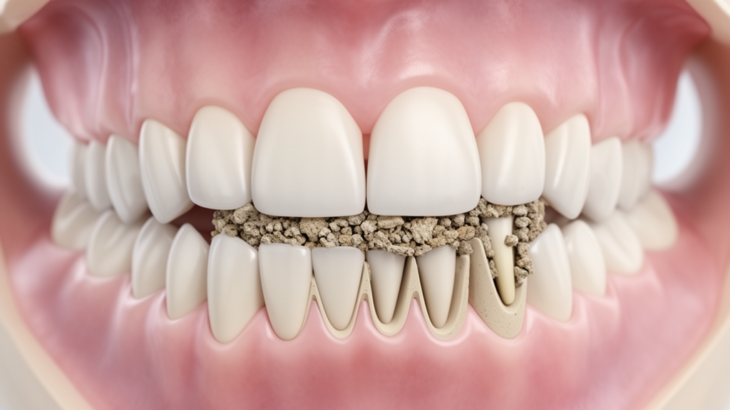
In implant dentistry, surgical reconstruction of deficient ridge areas within the anterior maxilla frequently exposes neighboring natural teeth to unanticipated structural resorption. A retrospective cohort clinical investigation published in Head & Face Medicine reveals that modifying standard guided bone regeneration protocols by deliberately extending particulate graft material across the facial plate of neighboring teeth counteracts crestal bone loss following full-thickness mucoperiosteal flap elevation. Conducted at the Affiliated Stomatology Hospital of Guangzhou Medical University, the study tracked 80 dental implant patients who contributed 135 adjacent natural teeth, demonstrating that covering the labial alveolar wall up to the facial alveolar crest effectively stabilized horizontal bone dimensions and produced measurable vertical dimensional gains over a six-month radiographic observation window.
The anatomical architecture of the anterior maxillary region presents documented clinical vulnerabilities that complicate both staged and simultaneous implant procedures. Research indicates that the labial alveolar wall in the majority of maxillary incisors and canines possesses a baseline thickness of less than one millimeter, predisposing the cortical plate to fenestrations, dehiscences, and rapid osteoclastic degradation when exposed during surgery. While guided bone regeneration remains an established cornerstone technique for restoring volume inside edentulous receptor sites, the associated reflection of full-thickness mucoperiosteal flaps and periosteal releasing incisions inevitably disrupts local periosteal microvascular networks. This disruption can trigger adverse bone remodeling across adjacent root surfaces, leading to gingival recession, interdental papillary collapse, and compromised long-term aesthetic outcomes.
To evaluate whether operative shielding could mitigate this localized bone reduction, researchers screened institutional surgical records spanning April 2018 through December 2024. The investigative cohort met rigorous inclusion parameters, requiring patients to be at least 18 years of age, categorized within American Society of Anesthesiologists physical status class I or II, and exhibiting periodontal health or well-controlled periodontitis with probing depths not exceeding three millimeters. All included surgical interventions involved simultaneous or staged implant fixtures placed within the maxillary incisor or canine zones, accompanied by full-thickness flap reflection bounded by two vertical releasing incisions situated one tooth beyond the defect margins. Individuals presenting with uncontrolled systemic disorders such as diabetes, recent antiresorptive therapy, heavy tobacco usage exceeding ten cigarettes per day, postoperative wound infections, or compromised radiographic imaging were systematically excluded.
The study classified adjacent teeth into three distinct comparative cohorts based on intraoperative coverage relative to the buccal bone wall. In the non-extended guided bone regeneration group, bone graft material was strictly confined to the primary edentulous defect without covering the adjacent natural tooth. In the partially extended group, graft material was extended across a portion of the adjacent buccal wall but stopped short of reaching the facial alveolar crest. In the fully extended guided bone regeneration group, surgeons systematically packed graft material over the entire exposed labial plate of the neighboring tooth, carrying the particulate layer coronally to completely cover the facial alveolar crest. All operative interventions were completed by two experienced oral surgeons employing an identical regenerative protocol consisting of deproteinized bovine bone mineral xenograft covered by a resorbable porcine collagen membrane extending at least two millimeters past defect borders.
Dimensional changes were measured utilizing three-dimensional cone beam computed tomography scans acquired immediately prior to surgery and at a six-month postoperative recall interval. Scans were captured with a standardized exposure protocol of 85 kilovolts, 4 milliamperes, a 6.5-second exposure duration, a 0.25-cubic-millimeter voxel dimension, and a 17-by-11-centimeter field of view. Using specialized planning software, pre- and postoperative digital imaging datasets were superimposed through a point-based rigid registration algorithm aligned to five fixed craniofacial hard-tissue reference landmarks, specifically the anterior nasal spine alongside paired zygomaticofacial and infraorbital foramina. This volumetric alignment achieved high registration accuracy, demonstrating an overall root mean square discrepancy of 0.13 millimeters across landmarks.
A calibrated examiner who demonstrated high intra-rater reliability identified the sagittal slice corresponding precisely to the longitudinal axis of each individual natural tooth. Primary outcome measures focused on linear horizontal thickness changes measured perpendicular to the tooth axis at the level of the facial alveolar crest, along with vertical displacement assessed through shifts in the distance separating the cementoenamel junction from the crestal margin. Secondary outcome parameters tracked horizontal wall dimensions measured at two, four, six, eight, and ten millimeters apical to the baseline crest, while additionally cataloging the proportion of vulnerable sites where initial bone thicknesses of one millimeter or greater deteriorated below the critical one-millimeter threshold following six months of postoperative remodeling.
Quantitative radiographic assessments revealed starkly divergent healing trajectories between treatment arms at the six-month evaluation. Teeth managed without graft extension sustained statistically significant crestal horizontal resorption averaging 0.40 ± 0.32 millimeters, while the partially extended approach experienced comparable horizontal reductions averaging 0.43 ± 0.44 millimeters. Conversely, adjacent teeth receiving the fully extended grafting protocol preserved horizontal dimensions, exhibiting a stable net change of 0.15 ± 0.75 millimeters. However, because the intra-rater reliability analysis established a 95 percent minimal detectable change threshold of 0.28 millimeters for horizontal linear measurements, the authors explicitly emphasized that this modest 0.15-millimeter positive shift cannot be reliably distinguished from standard technical measurement error.
Parallel patterns were recorded across vertical crestal measurements. Neighboring teeth allocated to the non-extended and partially extended groups experienced significant vertical alveolar crest resorption, with the distance from the cementoenamel junction to the crest increasing by 0.09 ± 0.20 millimeters and 0.03 ± 0.05 millimeters, respectively. In marked contrast, sites treated with the fully extended technique achieved a statistically significant vertical dimensional gain, reflected by a mean reduction in the cementoenamel junction-to-crest distance of 0.43 ± 1.01 millimeters. This vertical augmentation exceeded the corresponding 0.25-millimeter minimal detectable change threshold for vertical dimensions, confirming true structural gain rather than analytical variance.
Secondary evaluations assessing horizontal thickness across root levels demonstrated that non-extended and partially extended protocols suffered dimensional loss localized predominantly within the coronal two millimeters. In contrast, the fully extended technique yielded progressive horizontal dimensional increases extending from the crest down to eight millimeters apically. When examined by tooth category within the fully extended cohort, lateral incisors demonstrated the greatest horizontal gains, followed sequentially by canines and central incisors. Crucially, categorical assessment revealed that among teeth presenting with baseline facial wall thickness of one millimeter or greater, 100 percent in the non-extended group and 79.4 percent in the partially extended group thinned to less than one millimeter postoperatively, compared to only 33.3 percent in the fully extended group.
To adjust for patient-level clustering among individuals contributing multiple adjacent teeth, multivariable generalized linear mixed models were executed utilizing patient identification numbers as random intercepts. After adjusting for confounding covariates including patient age, sex, tobacco use, baseline periodontal phenotype, implant placement timing, and follow-up duration, surgical group allocation persisted as the primary significant predictor of post-surgical dimensional shifts. Compared with the non-extended reference standard, the fully extended intervention significantly predicted enhanced preservation of horizontal thickness with a regression coefficient of 0.48 and superior vertical crestal retention with a matching coefficient of 0.48, whereas the partially extended technique demonstrated no statistically significant advantage over non-extended treatment.
The study authors proposed several biological mechanisms explaining why full coronal graft coverage confers superior structural protection compared to incomplete or localized grafting. Applying a slowly resorbing xenogeneic scaffold beneath a collagen membrane creates a stable mechanical buffer that prevents periosteal soft-tissue collapse against denuded root surfaces during healing. Furthermore, completely shielding the fragile facial alveolar crest shields bare bone from aggressive osteoclastic resorption mediated by surgical trauma and inflammatory cascades. Because partial extension failed to prevent crestal recession, the authors noted that graft materials must actively encapsulate the crestal margin to neutralize the intense contractile and resorptive pressures exerted by the healing mucoperiosteal flap.
Despite these favorable findings, the investigators highlighted meaningful methodological limitations inherent to their retrospective cohort design. Patients were not randomly assigned to surgical groups, leaving open the possibility of surgeon selection bias based on initial clinical presentations. Furthermore, cone beam computed tomography cannot histologically distinguish newly formed viable bone from unresorbed particulate xenograft particles embedded in connective tissue. The six-month postoperative follow-up also remains insufficient to evaluate the multi-year structural and aesthetic stability of the augmented alveolar wall. The researchers concluded that fully extended guided bone regeneration covering the adjacent labial alveolar wall was associated with better preservation of radiographic ridge dimensions six months post-surgery than non-extended or partially extended techniques, although horizontal change remained below the minimal detectable change threshold.
Subject of Research: Radiographic preservation of adjacent alveolar bone dimensions using extended guided bone regeneration
Article Title: Effects of extended guided bone regeneration on the labial alveolar wall of adjacent teeth in the anterior maxilla: a retrospective cohort study
Article References: Zeng, X., Chen, H., Jiang, Y., Wen, Y., Luo, J., Chen, F., Chen, B., Zhao, S., & Xi, T. (2026). Effects of extended guided bone regeneration on the labial alveolar wall of adjacent teeth in the anterior maxilla: a retrospective cohort study. Head & Face Medicine, 22(1), Article 56. https://doi.org/10.1186/s13005-026-00624-y
Image Credits: AI Generated
DOI: 10.1186/s13005-026-00624-y
Keywords: guided bone regeneration, labial alveolar wall, anterior maxilla, dental implants, bone resorption, cone beam computed tomography, alveolar crest, facial bone thickness, periodontal bone support, Bio-Oss, Bio-Gide, cementoenamel junction, retrospective cohort study, implant dentistry, mucoperiosteal flap, generalized linear mixed models, aesthetic zone

