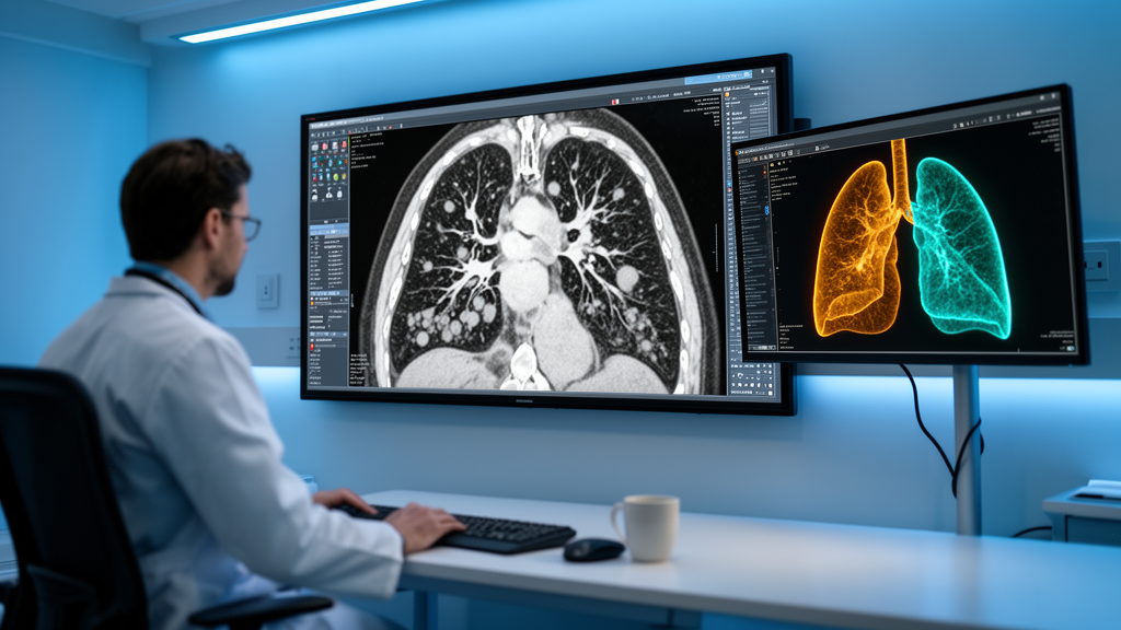
For thousands of patients with non-small cell lung cancer, targeted drugs called EGFR tyrosine kinase inhibitors represent one of modern oncology’s genuine success stories, shrinking tumors and extending lives with remarkable precision. Yet the victory is often temporary. Many tumors eventually acquire a specific genetic change known as the T790M mutation, which allows the cancer to shrug off the medication and resume its deadly advance. A new study from Zhejiang Cancer Hospital in Hangzhou, China, published in BMC Medical Imaging, suggests that the answer to which patients will develop this resistance may already be hiding in plain sight, inside the routine CT scans taken before treatment even begins.
The research team, led by Xianglan Zhou and Lei Shi of the hospital’s radiology department, developed a machine learning approach that combines two types of information: standard clinical characteristics and a technique called delta-radiomics, which captures how the texture and appearance of a tumor changes over time on imaging. Their goal was ambitious but practical. Rather than waiting for a tumor to progress and then performing an invasive biopsy to test for the T790M mutation, they wanted to predict the risk of that mutation emerging before first-line EGFR-TKI therapy was ever started.
The study included 285 patients with non-small cell lung cancer whose disease had progressed after first-line treatment with an EGFR tyrosine kinase inhibitor between January 2013 and September 2019. These patients were randomly divided into a training cohort of 213 individuals and a separate test cohort of 72, allowing the researchers to build their predictive models on one group and evaluate them honestly on another. Four distinct models were constructed and compared: one based purely on clinical characteristics, one on radiomics features extracted from non-contrast-enhanced CT scans, one on delta-radiomics features, and a fourth that combined clinical data with delta-radiomics.
Radiomics itself is a rapidly growing field that converts medical images into vast arrays of quantitative data. Rather than simply measuring the size of a tumor, radiomic algorithms extract hundreds of mathematical descriptors that characterize texture, shape, intensity patterns, and spatial relationships within the imaged tissue. Features derived from grey-level co-occurrence matrices, grey-level run-length matrices, and grey-level dependence matrices capture subtle patterns of pixel intensity that the human eye cannot perceive but that may reflect underlying biological properties such as cell density, necrosis, and vascular structure.
What makes delta-radiomics particularly powerful is its temporal dimension. Instead of analyzing a single scan, it computes the difference between radiomic features extracted from two time points, essentially quantifying how a tumor’s imaging phenotype evolves. This change over time may encode information about tumor heterogeneity and treatment response that a static snapshot cannot. In the context of this study, the delta approach allowed the machine learning models to detect imaging signatures associated with tumors that were biologically primed to develop the T790M resistance mechanism.
The results, measured on the independent test set, told a clear story. The clinical model alone achieved an area under the receiver operating characteristic curve, or AUC, of 0.70, with an accuracy of 66.7 percent. The NECT radiomics model performed worse, with an AUC of 0.63 and accuracy of 57.0 percent. The delta-radiomics model improved on both, reaching an AUC of 0.75 and accuracy of 72.2 percent. But the combined model, integrating clinical characteristics with delta-radiomics features, outperformed them all, achieving an AUC of 0.82 and an accuracy of 79.2 percent. Decision curve analysis, which evaluates the clinical net benefit of a predictive model across different threshold probabilities, further supported the combined model as the optimal choice.
One of the most significant contributions of this work lies not in the raw predictive numbers but in the effort to make the machine’s reasoning transparent. The researchers applied the Shapley additive explanations framework, commonly abbreviated as SHAP, to visualize and interpret how the model arrived at its predictions. SHAP, rooted in cooperative game theory, assigns each input feature a contribution value for every individual prediction, revealing which factors pushed the model toward predicting the presence or absence of the T790M mutation. This interpretability is critical in medicine, where clinicians are understandably reluctant to trust black-box algorithms that cannot explain themselves.
Through the SHAP analysis, the team identified ten significant features driving the model’s decisions: two clinical features and eight radiomic features. While the specific identities of these features reflect the particular dataset and modeling choices of this study, the broader implication is that measurable imaging characteristics, particularly those that change over the course of treatment, carry genuine predictive signal about the evolutionary trajectory of the tumor’s resistance mechanisms. This finding aligns with a growing body of evidence suggesting that the radiographic appearance of a tumor is a window into its genomic and microenvironmental state.
The clinical implications, if validated, could be substantial. Patients identified as high risk for developing the T790M mutation might be candidates for closer surveillance, earlier liquid biopsy testing, or alternative treatment sequencing strategies. Conversely, those predicted to be at low risk might avoid unnecessary interventions. The ability to anticipate resistance rather than react to it represents a meaningful shift in the management of EGFR-mutant lung cancer, where the emergence of T790M has historically been discovered only after disease progression has already occurred.
The authors are careful to note that their work represents a preliminary step rather than a finished clinical tool. The retrospective design, the single-center cohort, and the modest size of the test set all warrant caution, and the researchers explicitly state that further prospective multicentre validation is needed before the model can be applied in clinical practice. Nevertheless, the study demonstrates that the fusion of temporal imaging data with clinical variables, interpreted through explainable artificial intelligence, offers a viable path toward anticipating one of the most consequential resistance mechanisms in lung cancer. As radiomics and machine learning continue to mature, the humble CT scan may become not just a diagnostic record but a predictive instrument, reading the future of a tumor before that future arrives.
Subject of Research: Predicting acquired EGFR T790M resistance mutations in non-small cell lung cancer using delta-radiomics and clinical features with SHAP interpretability
Article Title: Predicting acquired T790M mutations in non-small cell lung cancer after EGFR-TKI therapy: integration of delta-radiomics and clinical characteristics with SHAP interpretation
Article References: Zhou, X., Shi, L., Lin, C., Zou, M., Sun, J., Song, Z., Wang, W., Ren, H., & Shi, L. (2026). Predicting acquired T790M mutations in non-small cell lung cancer after EGFR-TKI therapy: integration of delta-radiomics and clinical characteristics with SHAP interpretation. BMC Medical Imaging. https://doi.org/10.1186/s12880-026-02748-8
Image Credits: AI Generated
DOI: 10.1186/s12880-026-02748-8
Keywords: non-small cell lung cancer, EGFR-TKI, T790M mutation, delta-radiomics, machine learning, SHAP, computed tomography, drug resistance, predictive biomarkers, tumor heterogeneity, radiomics, precision oncology