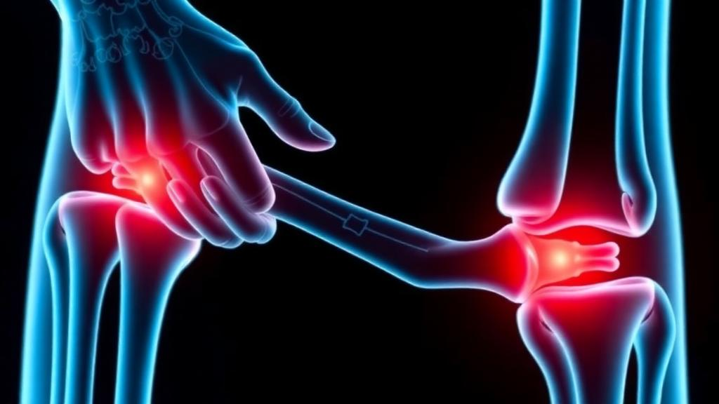
Triclosan, the synthetic antimicrobial that has quietly permeated everything from hand soaps and toothpastes to cutting boards and athletic wear, may be more biologically entangled with osteoarthritis than anyone suspected. A new integrative systems toxicology study published in iScience by Xijie Tang, Haijia Xu, Xiangzhong Liu, Yi Yang, and Zhanghua Li has assembled a multi-layered case that this ubiquitous environmental chemical, already flagged for endocrine disruption and immune dysregulation, appears capable of pushing cartilage cells toward the exact molecular behaviors that define joint degeneration. The work does not claim that triclosan causes osteoarthritis in people, but it maps, with unusual technical breadth, how the chemical’s known molecular fingerprints converge with the biology of a disease that afflicts hundreds of millions worldwide.
The team’s starting point was a recognition that osteoarthritis is no longer viewed as simple mechanical wear and tear. Contemporary research frames it as a chronic whole-joint disease involving the synovium, subchondral bone, and periarticular tissues, driven by inflammatory, immune, metabolic, vascular, and neuroendocrine processes. Environmental exposures such as phenols, phthalates, and particulate air pollution have emerged as potential modifiers of disease susceptibility, yet most reported exposure-disease links remain descriptive. What has been missing is a molecular bridge: a defined set of genes and pathways through which an environmental chemical could plausibly interact with the joint microenvironment. Building that bridge was the explicit goal of the new study.
The researchers began by mining publicly available transcriptomic data. They integrated three Gene Expression Omnibus datasets as a training cohort, corrected batch effects with the ComBat algorithm, and identified 1,154 differentially expressed genes between osteoarthritic and normal cartilage samples using strict statistical thresholds. In parallel, weighted gene co-expression network analysis uncovered thirteen gene modules, four of which correlated strongly with disease status. Merging the differential expression results with the disease-associated modules yielded a comprehensive set of 2,674 osteoarthritis-related genes. Separately, they compiled 122 predicted triclosan target genes from four independent chemical-protein interaction databases: SwissTargetPrediction, STITCH, ChEMBL, and the Similarity Ensemble Approach.
The intersection of these two gene sets produced just 30 shared candidate genes, and this compact overlap became the study’s analytical centerpiece. Functional enrichment analysis revealed that these genes cluster around biologically suggestive territory: prostaglandin metabolism, vascular regulation, oxidoreductase activity, cytokine binding, and pathways including arachidonic acid metabolism, NF-κB signaling, AGE-RAGE signaling, and calcium signaling. To separate signal from noise among the thirty candidates, the team constructed 113 predefined machine learning pipelines, combining twelve algorithms with different feature-selection strategies, and evaluated them through repeated stratified cross-validation plus three independent external cohorts. The winning model, a stepwise generalized linear model paired with elastic net regularization, achieved an area under the curve of 0.992 in training and maintained respectable performance of 0.727 to 0.873 across the external datasets.
Four genes survived this computational gauntlet: CBR3, MAOA, MMP13, and AVPR1A. Shapley additive explanations analysis, repeated across one hundred resampling iterations, identified CBR3 as the strongest and most stable contributor to model predictions, followed by MAOA and MMP13, with AVPR1A contributing more modestly. Expression comparisons confirmed that CBR3 and MMP13 were significantly upregulated in osteoarthritic samples while MAOA and AVPR1A were downregulated. Single-cell RNA sequencing of osteoarthritic cartilage added cellular texture to the picture: CBR3 was expressed broadly across seven annotated chondrocyte subpopulations, from homeostatic to hypertrophic and inflammatory chondrocytes, while MMP13 showed selective enrichment in pre-hypertrophic, fibrocartilage, effector, and hypertrophic populations. MAOA and AVPR1A appeared only sparsely at the single-cell level, hinting at more specialized roles.
Immune infiltration analysis using CIBERSORT deepened the systems view. Osteoarthritic samples showed significantly altered composition of memory B cells, eosinophils, macrophage subsets, mast cells, natural killer cells, plasma cells, and T cell subsets compared with normal controls. Correlation analysis revealed that CBR3, in particular, was associated with multiple immune cell populations, suggesting the candidate genes may participate in or mark immune-stromal programs active in diseased joints. The authors are careful to note that these are associative findings, since the public datasets contained no individual triclosan exposure measurements, but they extend the framework toward the whole-joint, immune-inclusive model of osteoarthritis that modern rheumatology increasingly embraces.
Structural biology then entered the picture. Molecular docking predicted that triclosan fits into the binding pockets of all four candidate proteins with favorable energies ranging from −6.2 to −7.6 kcal/mol, with the strongest predicted interactions for MAOA and MMP13. One-hundred-nanosecond molecular dynamics simulations confirmed that the docked complexes remained conformationally stable, sampling well-defined low-energy states with dynamic, intermittent hydrogen bonding between the chemical and its putative targets. The authors emphasize that computational binding compatibility is not proof of intracellular target engagement, but the simulations lend structural plausibility to the entire candidate set and provide a rational basis for the experiments that followed.
Those experiments, conducted in primary rat chondrocytes, delivered the study’s most striking results. After establishing that 7 micromolar triclosan preserved roughly 88 percent of cell viability, the researchers exposed chondrocytes to this low dose alone and in combination with interleukin-1β, the classic inflammatory cytokine of osteoarthritis. Triclosan alone elevated intracellular reactive oxygen species, and the combined treatment produced the strongest oxidative signal of all. Gene expression shifted in a selective, disease-relevant pattern: Cbr3 and Mmp13 rose, while Col2a1, the gene encoding the alpha chain of type II collagen, the very structural protein of cartilage, fell. Western blotting and immunofluorescence confirmed that CBR3 protein accumulated most strongly under combined treatment. Crucially, the response was not a blanket amplification of inflammation; Maoa and Avpr1a did not drop significantly below interleukin-1β-only levels, indicating a targeted redox and matrix effect rather than nonspecific cellular distress.
The biological logic that emerges is coherent. CBR3, a short-chain dehydrogenase/reductase regulated by the Nrf2 antioxidant pathway, appears to be the most consistent redox-responsive marker of the triclosan response. MMP13 is the key collagenase degrading type II collagen in osteoarthritic cartilage, so its upregulation alongside Col2a1 suppression signals a shift from matrix maintenance toward catabolic remodeling. MAOA, sitting on the mitochondrial outer membrane and generating hydrogen peroxide as a metabolic byproduct, points toward mitochondrial and autophagy-related vulnerabilities. The authors package these relationships into a hypothesis-generating adverse outcome pathway framework, explicitly exploratory, that connects chemical exposure to molecular initiating events, intermediate redox and inflammatory perturbations, and ultimately matrix degradation.
The limitations are candidly acknowledged. The human transcriptomic datasets contained no exposure measurements, the cell experiments used a single triclosan concentration over a short duration in rat rather than human cells, protein-level validation was restricted to CBR3, and no direct binding assays were performed. An NHANES-based study of urinary phenols previously found no significant association between triclosan and osteoarthritis prevalence, a fact the authors cite rather than avoid. What this study offers instead is a rigorous, testable map: future work should determine whether manipulating CBR3 or MAOA alters reactive oxygen accumulation, whether longer and dose-varied exposures reproduce the matrix-remodeling signature, and whether the predicted protein interactions hold up under direct experimental binding. For a chemical detected in the urine of most people surveyed, the possibility that it nudges cartilage cells toward catabolism is a hypothesis that now demands to be taken seriously.
Subject of Research: Molecular links between triclosan exposure and osteoarthritis investigated through integrative systems toxicology
Article Title: Potential molecular links between triclosan exposure and osteoarthritis: An integrative systems toxicology study
Article References: Tang, X., Xu, H., Liu, X., Yang, Y., & Li, Z. (2026). Potential molecular links between triclosan exposure and osteoarthritis: An integrative systems toxicology study. iScience, 29(11), Article 117868. https://doi.org/10.1016/j.isci.2026.117868
Image Credits: AI Generated
DOI: 10.1016/j.isci.2026.117868
Keywords: triclosan, osteoarthritis, systems toxicology, chondrocytes, reactive oxygen species, CBR3, MMP13, MAOA, machine learning, molecular docking, cartilage, environmental health