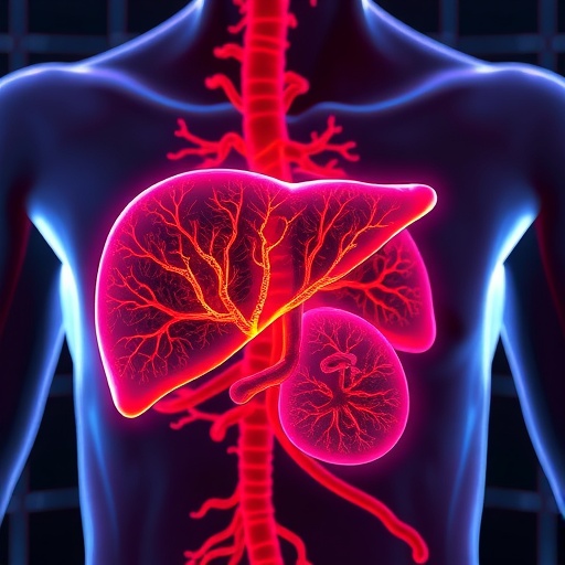
For decades, biologists have known that the liver is not a uniform chemical factory. Its cells perform different tasks depending on where they sit within the microscopic architecture of a liver lobule, the basic functional unit through which blood flows from the portal triad toward a central vein. Cells near incoming blood vessels specialize in some metabolic and detoxification activities, while cells closer to the central vein carry out others. This spatial pattern, known as liver zonation, allows one organ to divide potentially conflicting biochemical jobs with remarkable precision. A new study by Lee, Jakab, Uvarovskii and colleagues reports that this division of labor is controlled not only by hormones, nutrients and oxygen, but also by the physical forces generated by blood flow. Published in Nature Communications, the work describes a mechanism in which haemodynamics—the movement and pressure of blood—guides vascular Wnt signalling to instruct neighboring liver cells.
The finding offers a significant shift in the way scientists understand tissue organization. Liver zonation has traditionally been associated with gradients of oxygen, nutrients and metabolites established as blood travels through the lobule. Because oxygen levels are generally higher near the portal circulation and lower near the central vein, researchers have long proposed that oxygen availability helps determine which genes are active in each region. Yet oxygen alone cannot explain the full complexity of the liver’s spatial program. Many genes display sharply defined patterns even when their relationship to oxygen is indirect, and zonation can change during development, regeneration or disease. The new study places the blood vessels themselves at the center of this process, suggesting that they do more than deliver oxygen and remove waste: they actively send positional instructions to liver tissue.
At the heart of the mechanism is the Wnt signalling pathway, a communication system used throughout biology to control cell identity, growth and tissue renewal. Wnt proteins are secreted molecular signals that bind to receptors on nearby cells, regulating the stability of β-catenin and influencing the expression of genes that define cellular behavior. In the liver, Wnt activity has previously been linked to the identity of cells near the central vein, where it supports a characteristic set of metabolic functions. The new research indicates that Wnt signals produced or controlled by the vascular compartment are responsive to haemodynamic conditions. In other words, the endothelium—the thin layer of cells lining blood vessels—can interpret the physical environment created by flowing blood and translate it into a biochemical message for hepatocytes, the liver’s principal functional cells.
This concept is known as mechanochemical or mechanosensitive signalling. As blood moves through narrow vascular channels, it produces shear stress, a frictional force acting along the vessel wall, as well as pressure and changes in vessel shape. Endothelial cells possess molecular sensors capable of detecting these forces. Their response can alter intracellular calcium levels, transcriptional programs and the release of signalling molecules. According to the study, these haemodynamic cues influence vascular Wnt activity, establishing a local signal that helps maintain the functional identity of hepatocytes in different zones. The result is a feedback system in which blood flow is not simply a consequence of tissue architecture. Instead, flow becomes an instructive factor that helps create and preserve the architecture itself.
The implications are especially important because liver cells must coordinate dozens of specialized activities along a very short distance. Periportal hepatocytes, located near the entry of blood from the portal vein and hepatic artery, are generally associated with processes such as oxidative metabolism, urea production and certain aspects of gluconeogenesis. Pericentral hepatocytes, positioned closer to the central vein, are known for functions including xenobiotic processing, lipid metabolism and the expression of enzymes involved in drug transformation. These activities can generate toxic intermediates or require different oxygen conditions, making spatial separation essential. By linking local vascular signals to gene regulation, the mechanism described in the study provides a way for the liver to align metabolic programs with the exact physical environment experienced by each cell.
The researchers’ conclusions also help explain why disruption of blood flow can have effects that extend far beyond circulation. In conditions such as fibrosis, cirrhosis, portal hypertension and liver cancer, the normal geometry of vessels is remodeled. Scar tissue can compress or redirect vascular channels, while abnormal vessels may create regions with altered pressure, oxygenation and shear stress. If endothelial Wnt signalling depends on these physical variables, changes in blood flow could destabilize zonation even before widespread hepatocyte death occurs. The resulting molecular confusion may cause cells to activate inappropriate metabolic programs, increasing oxidative stress, impairing detoxification or making the tissue more vulnerable to injury. Zonation could therefore serve as both a functional adaptation and an early indicator of vascular pathology.
The study’s framework is also relevant to liver regeneration, one of the organ’s most remarkable biological abilities. After injury or surgical removal, surviving hepatocytes and supporting cells enter a coordinated repair program. Regeneration must restore not only the number of cells but also their spatial organization. A liver rebuilt without proper zonation might be structurally intact yet metabolically inefficient or dangerously prone to toxic reactions. If blood flow and vascular Wnt signals help re-establish positional identity, then the restoration of circulation may be an active component of regeneration rather than a final housekeeping step. This possibility could influence the design of organoids and engineered liver tissues, which often contain hepatocyte-like cells but struggle to reproduce the complex gradients and vascular relationships found in living organs.
The findings may also reshape how scientists approach drug testing. Many medicines are metabolized preferentially in particular liver zones, and toxic effects can emerge when compounds or their metabolites accumulate in the wrong cellular territory. Conventional liver cultures frequently lose their original zonation after removal from the body because they lack normal blood flow, vessel-derived cues and three-dimensional architecture. A culture system that reproduces haemodynamic forces and endothelial Wnt signalling could generate more realistic patterns of enzyme expression. Such models might improve predictions of drug toxicity, reveal why some compounds damage specific regions of the lobule and provide a more precise platform for testing therapies aimed at metabolic disease.
At the same time, the work does not suggest that Wnt signalling operates in isolation. Liver zonation is produced by the interaction of multiple gradients and cellular systems, including oxygen, nutrients, hormones, extracellular matrix composition, immune signals and nerve activity. The importance of the new study lies in identifying haemodynamics as an active component of that network and in connecting physical forces to a pathway already known to regulate liver identity. Further research will be needed to determine exactly how endothelial cells sense flow, which molecular intermediates connect mechanotransduction to Wnt production, and how quickly zonation changes when circulation is altered. Scientists will also need to establish whether the same mechanism operates across species and whether it can be safely manipulated without provoking uncontrolled cell growth, a recognized risk of excessive Wnt activity.
The broader message is that organs are shaped by conversations between chemistry and physics. In the liver, the route taken by blood appears to help tell each cell what kind of work it should perform. By revealing how haemodynamic information is converted into vascular Wnt signals, Lee and colleagues add a new layer to the biological map of liver function. The discovery could eventually guide treatments for vascular liver disease, improve regenerative medicine and make laboratory models more faithful to human physiology. For now, it offers a compelling explanation for how a constantly changing stream of blood can maintain one of the body’s most precisely organized metabolic landscapes.
Subject of Research: Haemodynamic regulation of liver zonation through vascular Wnt signalling
Article Title: Haemodynamic control of zonated liver function via instructive vascular Wnt signalling
Article References: Lee, K.H., Jakab, M., Uvarovskii, A. et al. Haemodynamic control of zonated liver function via instructive vascular Wnt signalling. Nature Communications 17, 8821 (2026). https://doi.org/10.1038/s41467-026-76966-7
Image Credits: AI Generated
DOI: https://doi.org/10.1038/s41467-026-76966-7
Keywords: liver zonation, haemodynamics, blood flow, vascular signalling, Wnt signalling, endothelial cells, hepatocytes, liver regeneration, liver disease, mechanotransduction