In a groundbreaking study set to transform our understanding of fibrotic skin disorders, researchers have uncovered a novel cellular interaction driving the abnormal bone-like tissue formation within keloids. This discovery hinges on the remarkable influence of catecholaminergic neurons, traditionally associated with neurotransmission, on fibroblast behavior. The team, led by Lou, Liang, Sun, and colleagues, presents compelling evidence that neural signals modulate osteogenic pathways in keloid fibroblasts, promoting their differentiation into bone-producing cells. Published in Nature Communications in 2026, this work pioneers a neurocutaneous axis in the pathology of keloid formation, challenging longstanding paradigms and opening new therapeutic avenues.
Keloids, notorious for their exuberant scar tissue extending beyond the boundaries of original wounds, have perplexed clinicians and researchers for decades. While excessive collagen production and aberrant fibroblast activity are well-documented hallmarks, the recent findings reveal a hitherto unappreciated dimension: the intrinsic osteogenic potential harbored within these fibroblasts, which is significantly augmented by signals derived from catecholaminergic neurons. These neurons synthesize catecholamines—dopamine, norepinephrine, and epinephrine—that appear to act in a paracrine fashion to prime fibroblasts toward bone formation, a phenomenon that aggravates the pathological rigidity of keloid tissues.
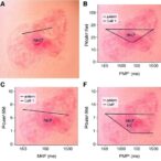
The investigation employed cutting-edge single-cell RNA sequencing and sophisticated neuroimaging techniques to delineate the cellular milieu within keloids. The researchers identified an enriched population of catecholaminergic neurons infiltrating the scar matrix, closely juxtaposed with fibroblasts exhibiting osteogenic gene signatures. Notably, the neurons exhibited elevated expression of enzymes responsible for catecholamine synthesis, suggesting a heightened neurochemical environment that supports fibroblast reprogramming. This spatial and molecular proximity underpins a functional neuro-fibroblast dialogue previously unrecognized in fibrotic skin diseases.
Crucially, functional assays reveal that catecholamines stimulate fibroblasts to produce key osteogenic markers including alkaline phosphatase, osteocalcin, and Runx2—a master transcription factor governing bone formation. The catecholamine-induced signaling cascades engage beta-adrenergic receptors on fibroblasts, activating downstream pathways such as cAMP/PKA and MAPK, which orchestrate the osteogenic transcriptional network. Pharmacological blockade of these receptors mitigated the osteogenic shift, underscoring the pivotal role of neuronal catecholamine signaling in this process.
Furthermore, in vitro co-cultures of catecholaminergic neurons with patient-derived keloid fibroblasts replicated the osteogenic phenotype, confirming the causative relationship. This neural influence extended beyond mere stimulation, as neurons appeared to sustain a microenvironment favoring fibroblast survival and pathological differentiation. The interplay culminated in the deposition of mineralized matrix components within the fibrotic scar, effectively mimicking bone tissue architecture and contributing to the characteristic rigidity and volume of keloids.
Animal models engineered to express increased catecholaminergic innervation in wounded skin mirrored the human condition, developing exaggerated keloid-like scars rich in osteogenic fibroblasts. Conversely, genetic or pharmacological ablation of these neurons resulted in attenuated scar fibrosis and diminished ectopic bone formation. These in vivo findings reinforced the concept that neural inputs are integral drivers of keloid pathophysiology and could represent a novel target for therapeutic intervention.
The implications of this work resonate across multiple disciplines. By unveiling a neurogenic component to fibroblast osteogenic activity, the study extends beyond dermatology into regenerative medicine, neurobiology, and fibrosis research. It accentuates the role of neuro-immune-fibroblast interactions in tissue remodeling and positions catecholaminergic neurons as critical regulators of pathological bone formation outside the skeletal system. This new framework compels a reevaluation of nervous system contributions to fibrotic diseases far beyond keloids.
Current treatments for keloids—ranging from corticosteroid injections to laser therapy—focus largely on suppressing inflammation and fibroblast proliferation but fail to address the aberrant differentiation into osteoblast-like cells. The discovery of catecholamine-driven osteogenesis opens an untapped therapeutic window. Targeting the neuronal inputs or the downstream adrenergic signaling in fibroblasts could halt or reverse the rigid calcification that complicates keloid management. Early-stage drug screening efforts exploiting beta-adrenergic antagonists have yielded promising preclinical results, suggesting feasibility for clinical translation.
This research also casts light on the enigmatic phenomenon of ectopic ossification in non-skeletal tissues, a process observed in other pathological settings such as fibrodysplasia ossificans progressiva and vascular calcification. The neurogenic influence reported here could serve as a unifying mechanism, where neural inputs act as modulators of mesenchymal cell fate toward bone lineage under aberrant conditions. Understanding these pathways at a mechanistic level may drive innovation in tackling a spectrum of fibrotic and calcific disorders.
Beyond therapeutic angles, the study encourages the development of diagnostic tools incorporating neural and osteogenic biomarkers to stratify keloid severity and predict treatment responses. Imaging modalities capable of identifying catecholaminergic neuron density or activity in scar tissue could facilitate early intervention before irreversible fibrosis ensues. This precision medicine approach would refine patient care and reduce the burden of chronic, disfiguring scars.
Moreover, the identification of a catecholaminergic neuro-fibroblast axis enriches the conceptual landscape of skin biology. The skin, traditionally viewed as a passive barrier and immune organ, emerges here as an active neuroendocrine interface, where nerves not only mediate sensation but also directly influence connective tissue remodeling. This paradigm fosters interdisciplinary collaborations, integrating neuroscience concepts into dermatological research and clinical practice.
In conclusion, the elucidation of catecholaminergic neurons as potent enhancers of fibroblast osteogenic activity in keloid scars heralds a paradigm shift in understanding fibrotic skin disorders. The multifaceted interplay between the nervous system and mesenchymal cells orchestrates pathological tissue remodeling, with profound implications for diagnosis, treatment, and regenerative strategies. This pioneering study by Lou, Liang, Sun, and colleagues paves the way for targeted neuro-modulatory therapies aimed at mitigating the burden of keloids and related fibrotic diseases, promising a future where neural biology and scar management converge for transformative clinical outcomes.
Subject of Research: Fibroblast osteogenic activity modulation by catecholaminergic neurons in keloid pathology
Article Title: Catecholaminergic neurons boost fibroblast osteogenic activity in keloid
Article References:
LOU, F., LIANG, J., SUN, Y. et al. Catecholaminergic neurons boost fibroblast osteogenic activity in keloid.
Nat Commun (2026). https://doi.org/10.1038/s41467-026-72823-9
Image Credits: AI Generated
Tags: catecholamine signaling in scar formationcatecholaminergic neurons in keloid pathologydopamine influence on fibroblast behaviorepinephrine-mediated fibroblast modulationkeloid fibroblast bone differentiationneural regulation of fibroblast osteogenesisneurocutaneous axis in fibrotic skin disordersneuroimaging in fibrotic researchnorepinephrine role in keloid developmentnovel therapies targetingosteogenic pathways in keloidssingle-cell RNA sequencing of keloid tissue