
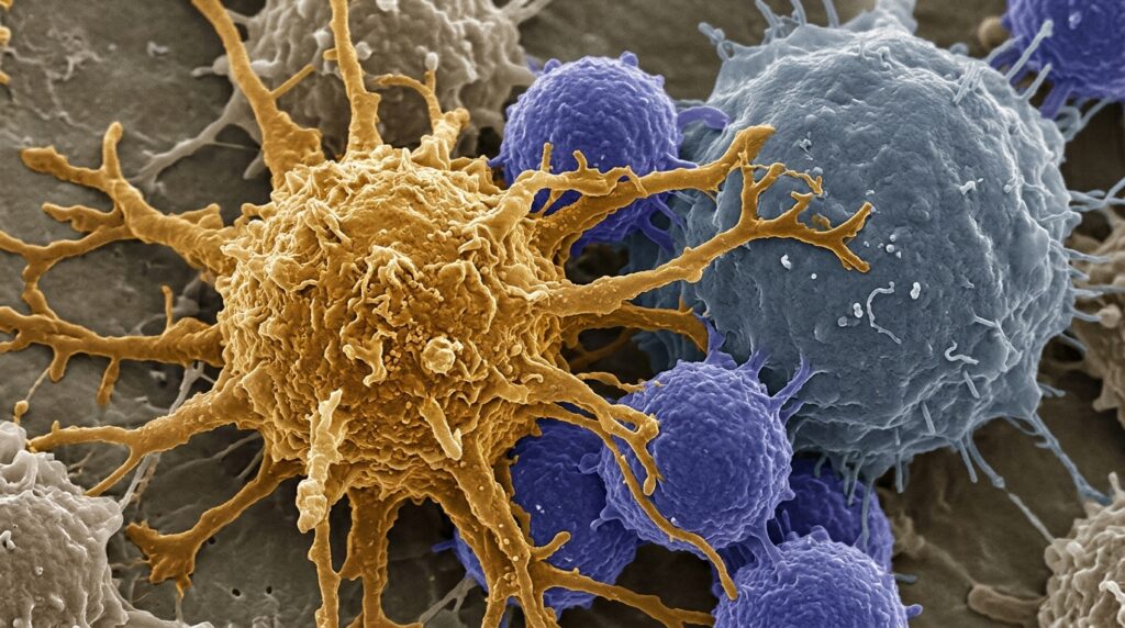
Dendritic cells enhance tebentafusp-mediated T cell activation, cytokine production, and cytotoxicity through a contact-dependent amplification mechanism, according to an experimental investigation published in Cancer Immunology, Immunotherapy. Tebentafusp represents the first approved T cell receptor-based bispecific therapy and prolongs overall survival in patients with metastatic uveal melanoma by redirecting functional T cells against tumor cells positive for gp100 and HLA-A*02:01. However, the precise ways in which this therapeutic agent reshapes broader immune-cell interactions within the surrounding tumor microenvironment have remained incompletely understood.
To investigate these immune dynamics, the research team employed HLA-defined donor T cells, dendritic cells, and distinct tumor cell models characterized by varied gp100 and HLA-A*02:01 expression profiles. Across the evaluated tumor cell models, the overall magnitude of tebentafusp-mediated functional responses showed a close association with both cellular gp100 abundance and HLA-A*02:01 surface expression. The experimental setup enabled researchers to systematically track how the addition of dendritic cells influenced bispecific-driven antitumor effector responses across varied antigen and receptor densities.
The observed dendritic cell-dependent functional enhancement operated independently of several established immune signaling routes. The authors determined that the amplification was not explained by conventional antigen presentation mechanisms or by the tested CD80/CD86 costimulatory pathway. Furthermore, the augmented T cell activation, cytokine production, and cytotoxic responses were not further enhanced when combining the treatment with immune checkpoint blockade targeting PD-1 or CTLA-4 pathways, demonstrating a distinct mode of action.
Direct cellular interactions proved essential for transferring tebentafusp onto dendritic cell surfaces. Following direct co-culture with tumor cells expressing both gp100 and HLA-A*02:01, even HLA-A*02:01-negative dendritic cells acquired and displayed surface-bound tebentafusp. These cross-decorated dendritic cells subsequently stimulated tebentafusp-specific T cell activation despite the underlying human leukocyte antigen mismatch between the dendritic cells and the target profiles.
Successful molecular transfer onto the dendritic cells required both tumor gp100 and HLA-A*02:01 expression alongside physical cell-to-cell contact, observations consistent with functional dendritic cell cross-dressing. Together, these laboratory findings identify a dendritic cell-mediated amplification mechanism that extends tebentafusp effector activity beyond direct tumor-T cell interactions, illustrating how accessory immune cells can actively reshape bispecific immune redirection within the local tumor microenvironment.
Subject of Research: Dendritic cell enhancement of tebentafusp-mediated T cell effector responses
Article Title: Dendritic cells enhance tebentafusp-mediated T cell effector responses
Article References: Wiesinger, M., Petzold, A., Luber, M., Hilgendorf, P., Wild, A., Heppt, M. V., Schuler-Thurner, B., Schober, K., Berking, C., & Koch, E. A. T. (2026). Dendritic cells enhance tebentafusp-mediated T cell effector responses. Cancer Immunology, Immunotherapy. https://doi.org/10.1007/s00262-026-04609-3
Image Credits: AI Generated
DOI: 10.1007/s00262-026-04609-3
Keywords: tebentafusp, dendritic cells, T cell activation, T cell receptor, bispecific therapy, uveal melanoma, gp100, HLA-A*02:01, cytokine production, cytotoxicity, cross-dressing, tumor microenvironment, antigen presentation, CD80/CD86 pathway, PD-1 blockade, CTLA-4 blockade, immune redirection