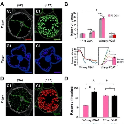
A new study published in Cell Death Discovery identifies diacylglycerol O-acyltransferase 1, or DGAT1, as an important regulator of macrophage pyroptosis during infection with Klebsiella pneumoniae. The findings place lipid metabolism at the center of an inflammatory process that can determine whether immune cells contain a bacterial invasion or contribute to the tissue damage associated with severe disease. The work, by HJ. Jung, SH. Jeong, SH. Lee and colleagues, adds to growing evidence that the immune response is shaped not only by microbial sensing, but also by the way cells store and process fats.
Klebsiella pneumoniae is a Gram-negative bacterium capable of causing pneumonia, bloodstream infections, urinary tract infections and life-threatening sepsis. Its prominent polysaccharide capsule can help it evade immune clearance, while strains carrying antimicrobial-resistance genes have become a major clinical concern. During infection, macrophages are among the first immune cells to recognize and engulf the bacteria. These cells release inflammatory signals and coordinate the recruitment of additional immune defenses, but excessive activation can damage surrounding tissues and intensify disease.
One of the most dramatic forms of inflammatory cell death is pyroptosis. Unlike the relatively quiet dismantling of a cell during apoptosis, pyroptosis causes the cell membrane to become permeable and eventually rupture. This process is commonly driven by inflammasome signaling, which activates inflammatory caspases and the pore-forming protein gasdermin D. The resulting membrane pores allow the release of cytokines such as interleukin-1 beta and interleukin-18, along with cellular contents that alert neighboring cells. Pyroptosis can help expose or eliminate intracellular pathogens, yet uncontrolled activation may amplify inflammation beyond the site of infection.
DGAT1 is best known as a metabolic enzyme. It catalyzes the final step in the synthesis of triacylglycerol, or triglyceride, by combining diacylglycerol with a fatty acyl-CoA molecule. This reaction supports the formation of lipid droplets, intracellular organelles that store excess fatty acids and provide a dynamic reservoir of energy and signaling molecules. Although lipid droplets were once considered passive fat depots, research now shows that they participate in immune signaling, organelle interactions and the response to infection. Altering DGAT1 activity can therefore influence both cellular metabolism and inflammatory behavior.
The new report links this metabolic pathway to the fate of macrophages exposed to K. pneumoniae. By identifying DGAT1 as a regulator of pyroptosis, the study suggests that the amount and organization of intracellular lipid may affect how macrophages respond to bacterial danger signals. Lipid metabolism could influence inflammasome assembly, the production of reactive molecules, mitochondrial stress or the availability of membrane components required for gasdermin-driven pore formation. These possible connections illustrate why infection biology increasingly views metabolism as an active part of immune regulation rather than a background process.
The relationship is especially significant in bacterial infections that provoke strong innate immune activation. Macrophages must balance two competing demands: they need to generate a rapid inflammatory response capable of restricting bacterial growth, while also preventing the response from destroying the tissue it is meant to protect. A regulatory factor such as DGAT1 could act within this balance by modifying the threshold for pyroptosis or changing the intensity of downstream inflammatory signaling. The precise direction and molecular sequence of the effect are central questions for interpreting the study and for determining whether DGAT1 might be therapeutically manipulated.
Potential treatments targeting this pathway would require considerable caution. Suppressing pyroptosis might reduce harmful inflammation, but it could also weaken a mechanism that helps the host detect and control bacteria. Conversely, enhancing inflammatory cell death might improve pathogen clearance in some circumstances while increasing the risk of lung injury, vascular leakage or systemic inflammation. The biological outcome would probably depend on the infectious strain, the tissue involved, the timing of intervention and the patient’s immune status. These factors make metabolic targets attractive but challenging candidates for drug development.
The findings also broaden the search for host-directed therapies against drug-resistant infections. Antibiotics act directly on bacterial growth or survival, whereas host-directed approaches seek to adjust the patient’s immune response or cellular environment. In principle, controlling DGAT1-associated signaling could complement antimicrobial treatment by limiting damaging inflammation without directly imposing additional selective pressure on the pathogen. However, such a strategy would need to preserve essential immune functions and avoid disrupting lipid balance in organs such as the liver, heart and adipose tissue, where DGAT1-related metabolism is also important.
Because the citation provides the study’s central conclusion but not its experimental details, the full article will be needed to determine how DGAT1 was manipulated, which macrophage models were used, and which molecular markers defined pyroptosis. Important questions include whether the investigators examined inflammasome components, inflammatory caspases, gasdermin D processing, cytokine release, bacterial burden or tissue pathology. Clarifying these points will show whether DGAT1 acts upstream of pyroptosis initiation, at the stage of membrane rupture, or through a broader metabolic program that influences several immune pathways at once.
The study’s central message is that the outcome of K. pneumoniae infection may depend on an intimate dialogue between microbial sensing, inflammatory cell death and lipid storage. By placing DGAT1 within that dialogue, Jung and colleagues highlight a potential connection between the chemistry of fat metabolism and the destructive power of innate immunity. The work does not make lipid metabolism a simple explanation for severe infection, but it offers a new framework for understanding why immune responses differ between patients and how future therapies might fine-tune inflammation rather than merely suppress it.
Subject of Research: DGAT1 regulation of macrophage pyroptosis during Klebsiella pneumoniae infection
Article Title: DGAT1 regulates macrophage pyroptosis during Klebsiella pneumoniae infection
Article References: Jung, HJ., Jeong, S.H., Lee, SH. et al. “DGAT1 regulates macrophage pyroptosis during Klebsiella pneumoniae infection.” Cell Death Discovery (2026). https://doi.org/10.1038/s41420-026-03272-y
Image Credits: AI Generated
DOI: https://doi.org/10.1038/s41420-026-03272-y
Keywords: DGAT1, macrophages, pyroptosis, Klebsiella pneumoniae, lipid metabolism, inflammasome, inflammation, bacterial infection, host-directed therapy
Tags: antimicrobial resistance in Klebsiella pneumoniaebacterial evasion strategiescellular fat storage and immune functionDGAT1 in macrophage pyroptosisimmune regulation during bacterial infectionKlebsiella pneumoniae infection mechanismslipid metabolism and immune responsemacrophage inflammatory cell deathpyroptosis in infectious diseasesregulation of inflammation by lipid enzymesrole of lipids in inflammationtissue damage from immune responses