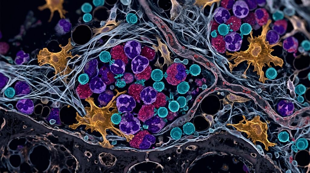
A comprehensive scientific review in Biomarker Research synthesizes evidence that the transformation of myelodysplastic neoplasms into acute myeloid leukemia is a dynamic ecological process driven by reciprocal interactions between mutant hematopoietic stem and progenitor cell clones and the bone marrow immune microenvironment. Although conventional models emphasize sequential genetic mutations as the primary driver of leukemogenesis, progression cannot be explained by mutational accumulation alone. Instead, abnormal hematopoietic clones evolve within a complex marrow niche comprising immune effectors, mesenchymal stromal cells, endothelial elements, and the extracellular matrix. Paired and serial sequencing studies reveal heterogeneous clonal trajectories in patients during leukemic progression, whereas most immunological and stromal investigations remain cross-sectional. Consequently, the review conceptualizes leukemic progression as an ecological continuum in which clonal evolution, chronic inflammation, immune exhaustion, stromal remodeling, and metabolic adaptation continuously reinforce one another, providing an integrated mechanistic framework rather than a descriptive catalog of isolated abnormalities.
In healthy bone marrow, balanced hematopoiesis and immune surveillance rely on coordinated signaling among hematopoietic progenitors, T cells, natural killer cells, monocytes, macrophages, dendritic cells, mesenchymal stromal cells, endothelial cells, and the extracellular matrix. Mesenchymal stromal cells maintain progenitor localization and self-renewal through CXCL12, stem-cell factor, adhesion molecules, and immunoregulatory factors, while CD8-positive cytotoxic T cells eliminate abnormal cells via T-cell receptor activation, cytotoxic granules, and cytokines. In early myelodysplastic neoplasms, this homeostatic balance shifts into a state where inflammatory activation coexists with impaired immune surveillance. Inhibitory checkpoint pathways including PD-1, TIM-3, and TIGIT constrain T-cell receptor signaling, reflecting pathway-level dysfunction rather than proven transformation triggers. Cross-sectional patient cohorts link elevated plasma ASC specks and aberrant cytokine profiles to disease progression, providing prognostic associations rather than causal proof. While marrow-restricted interleukin-6 signaling promotes leukemic progression in experimental mouse models, human data remain primarily cross-sectional and do not establish interleukin-6 as an obligate driver.
Chronic inflammation in the dysplastic marrow exerts selective pressure on hematopoietic clones while driving progressive genetic instability. Elevated levels of reactive oxygen species and reactive nitrogen species cause substantial oxidative and replication stress, inducing double-strand DNA breaks. Reduced function of DNA repair regulators such as MYBL2 impairs effective double-strand break repair, fostering subclonal diversity and providing the substrate for clonal selection. In the engineered NHD13 mouse model, PUMA-mediated apoptosis of progenitor cells actively accelerates leukemic transformation, demonstrating a causal link within that preclinical system rather than establishing a direct patient mechanism. Concurrently, inflammatory cell death pathways including necroptosis trigger extracellular release of high-mobility group box 1 protein, which engages toll-like receptors and stimulates persistent NF-κB signaling. Cross-sectional human marrow studies demonstrate that inflammatory stromal states correlate with diminished hematopoietic support, alongside altered distributions of granulocytic and monocytic populations, confirming that early disease involves continuous tissue injury and clonal adaptation rather than simple inflammatory elevation.
The erosion of adaptive and innate immune surveillance allows emerging preleukemic clones to escape destruction. In cross-sectional comparisons of patient bone marrow in myelodysplastic neoplasms and chronic myelomonocytic leukemia, CD8-positive T cells exhibit diminished repertoire diversity and pronounced oligoclonal expansion. Hypomethylating therapy remodels this repertoire, while combinations such as sabatolimab with hypomethylating agents expand cytotoxic natural killer and CD4-positive T cells, providing pharmacodynamic treatment evidence rather than tracking spontaneous transformation. High expression of PD-1 and TIM-3 on interferon-gamma-producing T cells correlates with adverse outcomes in acute myeloid leukemia cohorts, indicating exhaustion without proving a causal role in transformation. Concurrently, natural killer cell cytotoxicity is impaired despite altered major histocompatibility complex class I expression and stress ligands on clonal cells. Patient-derived mesenchymal stromal cells suppress natural killer cytotoxicity through TIGIT-related pathways, and Fc-gamma receptor IIIB deficiency impairs antibody-dependent cellular cytotoxicity, weakening the cooperation between innate and humoral defenses and reducing the clearance of abnormal hematopoietic clones.
Compromised cytotoxic immunity is reinforced by expanding immunosuppressive myeloid and regulatory populations that protect dysplastic clones from clearance. Patient bone marrow samples and functional assays show that myeloid-derived suppressor cells exploit the TIM-3 and Galectin-9 signaling pathway to inhibit T-cell function and promote exhaustion. Low-density granulocytes and abnormal monocyte subsets accumulate within the marrow, generating inflammatory mediators while suppressing T-cell and natural killer cell responses. Furthermore, hematopoietic clones harboring TP53 mutations alter cytokine secretion and immune-cell infiltration, and activated CD95-positive CD45RA-negative regulatory T cells associate with disease progression in discovery cohorts with clinical follow-up. Bone marrow macrophages also undergo significant functional reprogramming across risk groups, losing hematopoietic support capabilities and altering immunoregulatory programs. While thrombomodulin-expressing CD141-positive monocytes in lower-risk disease can dampen excessive immune activation, advancing disease is characterized by expanding suppressive myeloid networks that shield abnormal clones from immune surveillance and accelerate clinical progression.
Abnormal hematopoietic clones actively construct a protective niche through cell-intrinsic epigenetic and transcriptional alterations. In patient samples and cellular assays, EZH2-mediated trimethylation of histone H3 lysine 27 represses CXCL10 expression, reducing CD8-positive T-cell recruitment and driving functional exhaustion. Pharmacological inhibition of EZH2 in NHD13 mice restores CXCL10 expression and improves CD8-positive T-cell function, whereas SETD2 loss accelerates leukemogenesis in this model. Abnormal transcription factor activity also promotes clonal expansion; unmethylated RUNX1 enhances hematopoietic stem-cell survival and alters inflammatory gene expression in suppressive microenvironments. Spliceosome mutations further modify cellular fitness, with U2AF1-S34F linked experimentally to increased ribosome biogenesis and SF3B1 mutations driving distinct co-mutation-dependent progression routes. In human TP53-mutant myelodysplastic neoplasms, spatial profiling reveals localized DNA-damage-response neighborhoods, indicating that genomic instability and tissue architecture jointly foster transformation-permissive niches, although cross-sectional spatial data do not demonstrate whether these specialized neighborhoods precede or follow clonal expansion.
Mesenchymal stromal cells and extracellular matrix elements undergo extensive remodeling, disrupting physiological hematopoiesis while providing physical and biochemical protection to malignant clones. In mesenchymal stromal cells from young patients with excess blasts-1, loss of PD-L1 disrupts the SETDB1-H3K9me3 chromatin axis and induces a senescence-associated secretory phenotype with elevated interleukin-6, interleukin-8, and monocyte chemoattractant protein-1, creating a proinflammatory niche. Clonal TWIST1 expression in neoplastic myeloid cells promotes mesenchymal stromal cell differentiation toward adipogenesis and away from osteogenesis, degrading supportive niche structures. Preclinical models confirm that primary stromal defects can drive transformation independently; stroma-specific Setd2 deficiency in NHD13 mice accelerates leukemic progression and impairs hematopoietic support by upregulating coagulation factor XII. In addition, extracellular matrix remodeling characterized by increased hyaluronan deposition alters adhesion signaling, enhancing malignant cell survival, migration, and drug resistance. Histologically, altered niche organization manifests as abnormal localization and clustering of CD34-positive immature precursors escaping homeostatic niche constraints.
Metabolic reprogramming is an integral component of clone-stroma interactions that enables abnormal clones to adapt to hypoxic and inflammatory marrow conditions while fostering treatment resistance. Experimental studies show that GATA2-haploinsufficient hematopoietic progenitors display increased mitochondrial oxidative phosphorylation, reflecting distinct metabolic adaptations. In an experimental myelodysplastic mouse model, intracellular ASC specks strongly correlate with tissue iron deposition, and hepatocyte-targeted suppression of Tmprss6 increases endogenous hepcidin, reducing iron burden, dampening inflammasome activation, and prolonging survival. Furthermore, transcriptional regulators including GFI1B and BDH2 connect cellular metabolic state with stress responses, cell-cycle regulation, and differentiation. In vitro cellular assays indicate that aryl hydrocarbon receptor signaling reprograms malignant myeloid metabolism and modifies cytarabine sensitivity, identifying a candidate mechanism of niche-mediated drug resistance that requires patient-level validation. Epigenetic alterations within stromal cells, such as aberrant homeobox gene methylation, further modify stromal secretory programs. Together, these metabolic adaptations cooperate with immune and stromal remodeling to sustain clonal survival.
The transition from myelodysplasia to acute leukemia can be conceptualized as the progressive closure of three interacting positive-feedback circuits. The first circuit links chronic inflammation, reactive oxygen species-mediated DNA damage, clonal expansion, and persistent toll-like receptor and NF-κB signaling driven by high-mobility group box 1 release. The second circuit connects progressive T-cell exhaustion, immune-evasion pathways involving TIM-3, Galectin-9, and PD-1, increasing clonal burden, and intensified immune exhaustion. The third circuit links mesenchymal stromal abnormalities, metabolic support, and therapeutic resistance, sustained by altered stromal secretomes, adhesion signaling, and aryl hydrocarbon receptor activity. However, the authors caution that these circuits represent an integrative, hypothesis-generating synthesis assembled from independent cross-sectional patient cohorts, animal experiments, and cell-based assays. No single human study or animal model has demonstrated all components and transitions of either complete circuit, and selective pressures from treatments such as hypomethylating agents or stem-cell transplantation must be distinguished from the untreated natural history of disease.
Translating these microenvironmental insights into clinical practice requires rigorous validation before modifying patient care. Established risk assessment remains anchored in clinicopathologic systems including the revised and molecular International Prognostic Scoring Systems alongside morphology and cytogenetics; candidate microenvironmental biomarkers such as plasma ASC specks, cytokine profiles, CD95-positive CD45RA-negative regulatory T cells, or the research-stage Bcl-2:Ki-67 ratio require independent validation before altering routine risk stratification. Investigational therapies targeting the microenvironment, including checkpoint blockade against PD-1, TIM-3, or TIGIT, myeloid-derived suppressor cell inhibition, and NKG2D-directed chimeric antigen receptor T cells, demonstrate biological activity in early-phase trials but have not established clinical efficacy in preventing leukemic transformation. Future therapeutic strategies must shift toward coordinated multi-target interventions that combine clonal cytoreduction with immune restoration and stromal normalization. Validating these strategies will depend on prospective longitudinal studies using single-cell transcriptomics and spatial profiling across serial human bone marrow samples to identify reversible immune defects and establish truly individualized disease interception.
Subject of Research: Bone marrow immune microenvironment remodeling during myelodysplastic syndrome progression to acute myeloid leukemia
Article Title: The immune microenvironment in MDS-to-AML transformation: dynamic ecological remodeling and therapeutic implications
Article References: Long, Z., Wu, R., Yuan, S., Yang, K., Cheng, T., Han, B., & Yang, J. (2026). The immune microenvironment in MDS-to-AML transformation: dynamic ecological remodeling and therapeutic implications. Biomarker Research, 14(1), Article 167. https://doi.org/10.1186/s40364-026-01046-3
Image Credits: AI Generated
DOI: 10.1186/s40364-026-01046-3
Keywords: myelodysplastic neoplasms, acute myeloid leukemia, bone marrow microenvironment, hematopoietic stem cells, immune evasion, T-cell exhaustion, natural killer cells, myeloid-derived suppressor cells, mesenchymal stromal cells, chronic inflammation, leukemic transformation, extracellular matrix remodeling, metabolic reprogramming, immune checkpoints, TIM-3, PD-1, regulatory T cells