A retrospective clinical case series published in Maxillofacial Plastic and Reconstructive Surgery demonstrates the feasibility and spatial precision of combining robot-assisted surgery with a sandwich planning approach during complex mandibular reconstruction. Across all seventeen dental implants placed within transplanted bone flaps, the robotic platform achieved a mean longitudinal angular deviation of 1.17 degrees with a standard deviation of 0.30 degrees, while linear errors at both the implant entry points and apical endpoints remained strictly beneath half a millimeter.
Extensive mandibular defects frequently arise following radical resection of aggressive or malignant oral lesions, physical trauma, severe osteomyelitis, osteoradionecrosis, and medication-related osteonecrosis of the jaw. Reconstructive efforts must do more than simply bridge discontinuous skeletal segments; surgeons must simultaneously reestablish critical physiological mechanisms including mastication, deglutition, and articulate speech, all while preserving balanced facial aesthetics. Microvascular free flaps harvested from donor sites such as the fibula or the iliac crest represent the established standard of care for these challenging bony deficits. However, traditional multi-staged rehabilitation paradigms typically subject patients to multiple sequential operative interventions separated by prolonged recovery intervals. This extended surgical trajectory often leads to protracted periods of severe edentulism, progressive functional and aesthetic deterioration, compromised psychological quality of life, and compounded cumulative surgical morbidity accompanied by escalating healthcare expenditures.
To circumvent the drawbacks associated with staged reconstruction, surgical teams developed the jaw-in-a-day paradigm by combining virtual surgical planning, computer-aided design and computer-aided manufacturing technologies, and immediate fixed temporary dental restorations. This combined modality allows surgical teams to perform oncological bone ablation, harvest and shape microvascular bone flaps, drill implant osteotomies, insert endosseous implants, and seat rigid provisional prostheses within a single operational event. By eliminating months of interim healing without functional dentition, the protocol aims to restore immediate oral architecture and accelerate recovery. Nevertheless, conventional implementations rely heavily on pre-manufactured static 3D-printed surgical cutting and drilling guides. These static guide templates introduce significant positional variability stemming from physical guide plate positioning, manual drill instability, mismatched bone-plate adaptation, intervening soft tissue displacement, and individual operator variance, occasionally producing angular errors between two and four degrees alongside linear discrepancies of one to two millimeters.
To overcome the technical shortcomings of static mechanical templates, surgical robotics has entered reconstructive craniomaxillofacial procedures, offering continuous dynamic navigation, stereotaxic robotic arm guidance, and force-sensitive haptic boundary feedback. Robotic platforms directly interface three-dimensional digital plans with intraoperative tracking systems, maintaining real-time spatial awareness and robotic arm stability even within restricted anatomical boundaries. Previous experimental investigations indicated that robotic assistance could elevate osteotomy accuracy and anatomical boundary matching during fibular flap contouring beyond the tolerances of freehand or optical navigation methods. However, the comprehensive integration of active robotic systems within the full jaw-in-a-day workflow, specifically for coordinating microvascular tissue transfer, workbench implant site osteotomy, and instantaneous provisional prosthetic seating, had previously remained sparsely documented in peer-reviewed clinical literature.
The planning phase in this surgical cohort centered on a specialized protocol termed the sandwich approach, a computer-assisted design sequence in which engineers plan the definitive positions of the grafted bone segments and prosthetic dental arches prior to calculating the coordinates of the underlying dental implants. Preoperative imaging protocols involved head and neck contrast-enhanced computed tomography, cone-beam computed tomography, and contrast-enhanced donor-site computed tomography of the fibular or iliac vascular beds. Using ProPlan CMF software, surgical teams simulated tumor boundaries and manufactured patient-specific osteotomy guides, mirroring the unaffected contralateral mandibular contour to guide reconstructive geometry. Surgeons positioned the reconstructed bone fragments twelve to fifteen millimeters beneath the planned occlusal plane, securing sufficient vertical restorative space for prosthetics. Using 3Shape Implant Studio, clinicians established occlusion-driven implant vectors, depths, and diameters before transferring the combined models into Remebot robotic software to simulate instrument trajectories and prevent physical collisions.
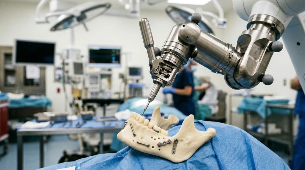
During surgery under general anesthesia, ablative surgeons first excised the mandibular tumors according to the predesigned resection guides, isolating recipient blood vessels in the neck while an adjacent team harvested the selected vascularized iliac or fibular flap with intact vascular pedicles. Rather than performing robotic drilling directly inside the patient’s oral cavity, the harvested bone grafts were mounted onto 3D-printed stereolithographic mandibular defect models resting on an adjacent in-vitro sterile workbench. After rigidly securing the vascularized bone segment to the reference model with mini-titanium plates, surgeons calibrated the optical tracking arrays and registered physical fiducial markers to align the real graft with the virtual plan.
Once all implants were anchored into the bone flap on the surgical workbench, clinicians applied a prefabricated 3D-printed positioning template to confirm the passive mechanical fit and spatial orientation of the provisional dental prosthesis. Surgeons then transferred this composite unit directly into the mandibular defect site, reestablishing systemic perfusion by performing microvascular anastomosis of donor and recipient blood vessels under an operative microscope. The bony segments were anchored to the residual mandibular stumps using pre-bent titanium plates, and fixed non-functional provisional prostheses were secured. Clinicians adjusted the artificial teeth out of occlusion to prevent disruptive masticatory forces during early osseointegration, maintaining intermaxillary elastic traction for four weeks postoperatively to stabilize alignment.
The clinical series evaluated five individual reconstructive operations involving varied anatomical locations, flap selections, and pathological tumor diagnoses. Case one involved a 36-year-old female presenting with an ossifying fibroma spanning the left mandibular body and ascending ramus, successfully repaired using a double-barrel fibula flap harboring two simultaneously placed implants supporting a functional prosthesis through six months of follow-up. Case two treated a 54-year-old female diagnosed with an odontogenic myxofibroma extending across teeth forty-three to thirty-seven, reconstructed using a two-segment double-barrel fibular flap with four implants, receiving a fixed provisional prosthesis at post-surgical week three.
The remaining three patients received vascularized iliac crest bone flaps tailored to their respective mandibular resections. Case three involved a 51-year-old female with a right posterior mandibular lesion spanning teeth forty-three through forty-seven, where a single-segment iliac flap supported three concurrent implants; although preoperative biopsy indicated benign ameloblastoma, definitive postoperative histopathology revealed clear cell odontogenic carcinoma, requiring intensive oncological surveillance that verified no tumor recurrence over twenty-two months. Case four treated a 43-year-old female with an extensive ameloblastoma from tooth forty-three to thirty-six using a two-segment iliac flap and four dental implants. Case five addressed a giant cell granuloma spanning teeth forty-three to forty-seven in a 38-year-old male using a single-segment iliac flap with four implants. All three iliac cases demonstrated stable bone healing and aligned dentition at six-month follow-up.
To rigorously quantify placement accuracy, investigators captured postoperative cone-beam computed tomography scans and superimposed them onto preoperative virtual surgical plans using the robotic system’s integrated verification module. The software registered the volumetric datasets using a semi-automated point-cloud matching algorithm, calculating three-dimensional vector deviations at the implant entry point, apical tip, and along the central longitudinal axis. Two independent investigators repeated each calculation twice to minimize manual measurement bias. Analysis of all seventeen placed implants revealed a mean global angular deviation of 1.17 degrees with a standard deviation of 0.30 degrees. The mean spatial deviation was 0.26 millimeters with a standard deviation of 0.09 millimeters at the entry point, and 0.24 millimeters with a standard deviation of 0.08 millimeters at the apical tip, fully satisfying predefined safety thresholds of two degrees and 0.5 millimeters.
In evaluating the clinical utility of the robotic platform, the authors noted that performing implant site preparation on an external workbench eliminated oral optical constraints and soft tissue interference, an environment where static guides could theoretically provide comparable precision at lower equipment cost. Balanced against these capabilities are substantial operational trade-offs, including high initial equipment acquisition costs, extended technical learning curves for multidisciplinary surgical staff, and prolonged operating and anesthesia durations necessitated by hardware setup, optical registration, and system calibration.
The authors emphasized several fundamental methodological limitations inherent to their preliminary investigation, pointing out that the retrospective design, small sample size of five patients, and absence of a concurrent parallel control group preclude claims of clinical superiority over standard guided methods. Furthermore, the six-month follow-up horizon provides an evaluation of early technical feasibility and initial implant stability, but cannot assess long-term biological outcomes such as peri-implant marginal bone loss or late mechanical stability. The investigators concluded that while combining robotic surgical navigation with the sandwich planning methodology represents a feasible strategy for achieving sub-millimeter dental implant placement in complex vascularized mandibular flaps, multicenter prospective randomized controlled trials with extended follow-up periods are required to establish its definitive clinical role.
Subject of Research: Feasibility and implant accuracy of robot-assisted jaw-in-a-day mandibular reconstruction using sandwich planning
Article Title: Robot-assisted jaw-in-a-day based on the sandwich approach: case series
Article References: Luo, P., Gao, X., Su, L., Chen, H., Deng, J., Ren, X., Fu, G., Huang, Y., Ou, Z., & Zhang, F. (2026). Robot-assisted jaw-in-a-day based on the sandwich approach: case series. Maxillofacial Plastic and Reconstructive Surgery, 48(1), Article 15. https://doi.org/10.1186/s40902-026-00513-z
Image Credits: AI Generated
DOI: 10.1186/s40902-026-00513-z
Keywords: jaw-in-a-day, mandibular reconstruction, robot-assisted surgery, sandwich planning, dental implants, virtual surgical planning, fibular flap, iliac flap, stereolithography, microvascular anastomosis, cone-beam computed tomography, osseointegration, point-cloud matching, ameloblastoma, computer-aided manufacturing, surgical accuracy