
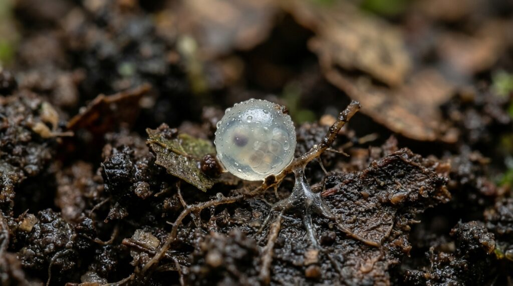
Developmental biologists studying the eyeless soil-dwelling mite Archegozetes longisetosus have discovered that its developing embryos transiently initiate molecular cascades characteristic of eye specification, uncovering hidden vestigial eye primordia within an organism that completely lacks adult visual organs. In animals across disparate metazoan lineages, the formation of visual structures traditionally relies on the conserved transcription factor Pax6 interacting with a coordinated group of co-factors collectively termed the retinal determination gene network. However, recent developmental evidence in Chelicerata, the arthropod group comprising arachnids, horseshoe crabs, and sea spiders, suggested that Pax6 orthologs do not direct ocular morphogenesis in the same manner observed in mandibulate arthropods. When genomic sequencing of A. longisetosus demonstrated that the eyeless arachnid retains two distinct Pax6 paralogs known as eyeless and twin of eyeless, along with single-copy orthologs of retinal determination network genes, researchers hypothesized that these sequences persisted either to drive non-visual embryonic patterning or to construct fleeting vestigial ocular tissues.
To investigate these developmental mechanisms, researchers Isabella Joyce and Austen A. Barnett examined the temporal and spatial distribution of retinal determination and head-patterning transcripts throughout embryogenesis in A. longisetosus. Because adult females of this species maintain oviducts that serve as internal brood chambers, laid egg clutches routinely contain embryos situated at varying developmental stages, preventing the standard use of hours after egg laying as a chronological metric. In response, the authors formulated a staging system dedicated to head and brain morphogenesis, establishing standardized stages from early germ-band stages through Brain Development Stage 1 up to Brain Development Stage 7 and the subsequent prelarval stage. By utilizing modern confocal microscopy paired with single and double hybridization chain reactions, the investigation generated a detailed spatial description of mite cephalic development, permitting direct morphological and molecular comparisons with eyed chelicerate exemplars such as wandering spiders and daddy-longlegs.
Morphological analysis of embryonic development demonstrated that the brain of A. longisetosus arises from the anterior pre-cheliceral ocular region, an anterior embryonic domain characterized by the early expression of Six3 and orthodenticle. During Brain Development Stage 1, this region bifurcates into bilateral optic lobes, which quickly develop distinct invaginating neural pits and paired anterior and lateral furrows at Brain Development Stage 2. As development progresses through intermediate stages, the anterior and lateral furrows deepen and, by Brain Development Stage 5, coalesce into a single continuous groove, a morphogenetic movement not documented in spiders or harvestmen. Cells forming the medial subdivisions then extend across these continuous channels, partitioning the neuroectodermal furrows into the arcuate bodies, mushroom bodies, and paired optic vesicles beneath the migrating non-neural prosomal shield. In the post-embryonic prelarval stage, the entire cephalic complex undergoes an inversion where the brain migrates dorsally, placing the arcuate bodies and associated structures in an upside-down orientation relative to earlier embryonic stages.
To evaluate whether terminal visual structures form during embryonic development, the authors evaluated transcripts responsible for phototransduction and photoreceptor differentiation. In eyed arachnids and related arthropods, the visual pigment rhodopsin, the non-visual opsin peropsin, and regulatory beta-arrestins mark mature and rudimentary visual organs. Hybridization chain reaction assays targeting the sole opsins preserved in the mite genome, Al-peropsin and Al-rhodopsin-7, revealed an absolute absence of transcription within the developing ocular region at every stage examined. Similarly, tests targeting three candidate beta-arrestin orthologs identified from transcriptomic libraries showed no detectable signal in the cephalic domain, while phylogenetic searches failed to identify any ortholog for the photoreceptor-associated structural motor protein myosin-III, known as ninaC in fruit flies. The systematic absence of these terminal visual markers verified that A. longisetosus halts visual morphogenesis long before differentiating functional photoreceptors or terminal optic tissues.
In contrast to the absence of terminal photoreceptor genes, upstream components of the retinal determination gene network exhibited clear, localized transcription in the ocular margins during early embryogenesis. The single-copy ortholog of eyes absent, designated Al-eya, appeared during early segmental stages in the lateral ocular region and medial groove ectoderm before expanding into bilateral patches lining the lateral furrows at Brain Development Stage 2. By intermediate stages, Al-eya marked both neural furrows and the peripheral ectoderm of the head lobes, precisely paralleling the pre-retinal domains found in eye-bearing spiders. Likewise, the transcriptional co-factor Al-sine oculis became active at Brain Development Stage 2 within the medial groove and the lateral margins of the head lobes, expanding to line the rim of each ocular lobe before being completely silenced across the cephalic domain by Brain Development Stage 7, consistent with the transient specification and subsequent suppression of vestigial eye fields.
Further tracking of retinal determination network orthologs illustrated clear differences in how individual genes are deployed during mite cephalogenesis. The transcription factor Al-dachshund showed no expression in the embryonic ocular region across early or intermediate stages, appearing in cephalic tissues only during the post-embryonic prelarval stage within the mushroom bodies and arcuate bodies, suggesting that downstream activation by eyes absent and sine oculis is decoupled or repressed in this lineage. The homeobox gene Al-Six3 demonstrated early transcription across the anterior pole and remained confined to neuroectodermal structures, including the developing mushroom bodies, lateral furrows, optic vesicles, and the compact ventral synganglion of prelarvae, with no signal in the peripheral non-neural ectoderm. Meanwhile, the basic helix-loop-helix factor Al-atonal displayed fleeting expression in bilateral cell clusters flanking the lateral furrows at Brain Development Stage 2, disappearing entirely from the ocular territory in subsequent stages.
Detailed localization of the Pax6 ortholog Al-eyeless indicated that this master regulatory gene functions predominantly in assembling the central nervous system rather than specifying visual organs. Earliest transcripts of Al-ey appeared during prosomal segmentation as paired triangular fields in the pre-cheliceral region overlapping an anterior stripe of Al-wingless, alongside segmental clusters within the nascent ventral nerve cord. As brain morphogenesis proceeded, Al-ey expression cleared from the floor of the lateral furrows, forming a ring-like and eventually C-shaped perimeter surrounding the invaginating furrows. In deeper focal planes, Al-ey-positive cells internalized into the cephalic interior to form defined components of the optic vesicles, mushroom bodies, and arcuate bodies. Segmental expression persisted through prelarval stages into the brain and ventral nerve cord, but Al-ey was entirely absent from the non-neural surface ectoderm where arachnid eye lenses and retinas develop.
The second Pax6 paralog, Al-twin of eyeless, displayed dynamic and divergent spatial kinetics that separated its role from both Al-ey and ancestral eye-specification programs. Initially transcribed across a broad diagonal band encompassing the cheliceral segment and anterior head region where it overlapped with Al-ey and Al-wingless, Al-toy expression rapidly cleared from the ocular territory as limb buds emerged. The gene then exhibited appendicular expression at the anterior margins of the prosomal limbs before disappearing from head tissues during mid-embryogenesis. By Brain Development Stage 7, Al-toy was expressed ubiquitously throughout the migrating prosomal shield as this ectodermal tissue advanced over the developing brain, demonstrating a specialized structural role in shield morphogenesis that contrasts sharply with the developmental patterns documented in spiders.
Simultaneous detection of the conserved head-patterning homeobox gene Al-orthodenticle illuminated how ancestral regulatory genes coordinate early brain compartmentalization. In pre-segmental and early blastoderm embryos, Al-otd was transcribed across the anterior margin and incipient ventral nerve cord, co-expressing extensively with both Al-ey and Al-toy within the presumptive protocerebral neuroectoderm. During intermediate stages, Al-otd marked cell blocks flanking the stomodaeum and the lateral furrows, establishing defined spatial boundaries with Al-ey in the medial subdivisions and the lateral grooves. By Brain Development Stage 7, Al-otd transcription shifted into the margins of the fused prosomal shield while maintaining ventral nerve cord expression, paralleling patterns documented for otd2 in spiders and otd in harvestmen. These synchronized expression dynamics indicate that Pax6 and orthodenticle cooperate to delineate the basal arachnid protocerebrum independently of any functional commitment to terminal retinal differentiation.
Taken together, the developmental findings demonstrate that Archegozetes longisetosus preserves morphological and transcriptional hallmarks of an ancestral visual past despite having secondarily lost its eyes in soil environments. The transient appearance and subsequent clearing of Al-eya and Al-so reveal that vestigial eye primordia are specified during early embryogenesis before being terminated prior to photoreceptor differentiation. Simultaneously, the persistence of lateral furrows, optic vesicles, and Pax6 expression reflects structural developmental constraints, wherein neural components of the visual pathway remain integrated into the architecture of the arcuate and mushroom bodies. The researchers emphasize that definitive verification of these gene functions and potential suppressive mechanisms, such as inhibitory Wnt signaling or evolutionary developmental burden, awaits the establishment of targeted genetic knockout or knockdown methodologies such as RNA interference and single-cell sequencing in acariform mites.
Subject of Research: Vestigial eye primordia and retinal determination gene expression in Archegozetes longisetosus
Article Title: The expression of Pax6 and retinal determination genes in the eyeless arachnid A. longisetosus reveals vestigial eye primordia
Article References: Joyce, I., & Barnett, A. A. (2025). The expression of Pax6 and retinal determination genes in the eyeless arachnid A. longisetosus reveals vestigial eye primordia. EvoDevo, 16(1), Article 12. https://doi.org/10.1186/s13227-025-00245-7
Image Credits: AI Generated
DOI: 10.1186/s13227-025-00245-7
Keywords: Archegozetes longisetosus, Pax6, Retinal Determination Gene Network, Arachnid Brain Development, Vestigial Eye Primordia, Hybridization Chain Reaction, EvoDevo, Acariform Mites, expression, retinal, determination, genes