
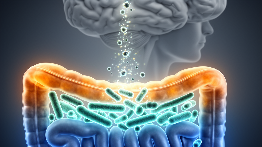
Parkinson’s disease has long been treated as a disorder of the brain, but a new open-access review published in Discover Immunity argues that some of its most important drivers may sit much lower in the body. The review, authored by Vaishnavi Suresh Jadhav, Deepak Nathiya, Balvir Singh Tomar and Dharmendra Kumar Khatri, synthesizes clinical, experimental and multi-omics evidence to build a unified picture in which gut microbial dysbiosis disrupts mucosal immunity, weakens the intestinal barrier, fuels systemic inflammation and ultimately leaves the nigrostriatal dopamine pathway vulnerable to degeneration. As a review, it integrates findings from many studies rather than reporting new experimental data of its own, and the authors are careful to note where the evidence remains associative.
The core of the argument rests on a recurring microbiome pattern. Across numerous cohorts, people with Parkinson’s disease show a marked depletion of short-chain fatty acid–producing commensals, including Faecalibacterium prausnitzii, Roseburia, Prevotella, Butyricicoccus and Blautia species, alongside an expansion of pro-inflammatory, endotoxin-enriched taxa such as Enterobacteriaceae, Akkermansia and Desulfovibrio. Short-chain fatty acids such as butyrate, propionate and acetate are the fermentation products of dietary fiber, and they carry anti-inflammatory, barrier-stabilizing and neuroprotective properties. When these taxa decline, tight junction integrity in the gut epithelium weakens, intestinal immune regulation falters, and microglial maturation and anti-inflammatory signaling in the brain are diminished.
The flip side of that loss is an excess of inflammatory triggers. Enriched taxa in Parkinson’s disease are associated with a heightened lipopolysaccharide burden, mucin degradation and increased reactive oxygen species generation. Elevated LPS activates TLR4-dependent inflammatory pathways, and clinical and experimental studies have linked raised plasma LPS and LPS-binding protein with disease severity and increased systemic cytokines. The resulting peripheral inflammatory state, characterized by elevated circulating IL-1β, IL-6, TNF-α and chemotactic mediators, is proposed to prime microglia, facilitate immune cell infiltration into the central nervous system through a weakened blood–brain barrier, and potentiate oxidative and mitochondrial stress within the substantia nigra.
The review also gives detailed attention to the alpha-synuclein connection. Accumulating findings suggest that intestinal inflammation may trigger misfolding of alpha-synuclein within enteric neurons, enabling its propagation to the brainstem via vagal pathways. This echoes Braak’s staging hypothesis, supported by postmortem and animal model evidence, which proposes that misfolded aggregates arise first in the enteric nervous system and olfactory bulb before spreading trans-synaptically to the midbrain. Epidemiological data add a provocative note: truncal vagotomy in humans has been associated in some large registry studies with lowered Parkinson’s risk, although other analyses and meta-analyses have found inconsistent associations, and the authors stress that vagal propagation may account for only a subset of cases.
Animal models supply much of the mechanistic weight. In the landmark work by Sampson and colleagues, alpha-synuclein–overexpressing mice raised germ-free showed reduced motor deficits, decreased microglial activation and attenuated alpha-synuclein pathology, while colonization with microbiota from human Parkinson’s donors restored motor impairment and neuroinflammation. Antibiotic depletion paradigms have largely reproduced these protective effects in MPTP- and alpha-synuclein-driven models. Yet the review highlights a striking complication: supplementing short-chain fatty acids to germ-free alpha-synuclein mice restores microglial maturation but also reinstates motor impairments and neuroinflammation, indicating context-dependent functions for these metabolites in disease models.
Human translational evidence is growing but uneven. Cross-sectional metagenomic and metabolomic studies across geographically diverse cohorts consistently report the depletion of butyrate-producing taxa and enrichment of mucin-degrading or pro-inflammatory genera, with some taxa correlated with constipation, disease duration and motor scores. Longitudinal and prodromal research, including cohorts with REM-sleep behavior disorder, suggests particular microbial signatures may exist before motor onset, hinting at a contributory rather than purely secondary role. Machine-learning classifiers trained on microbiome features reach average AUC values of roughly 0.70 to 0.75, a substantial but not conclusive disease signal, and models built on functional metagenomic pathways such as butyrate synthesis outperform taxonomy-only approaches. The authors caution that variability across studies, driven by diet, medications such as levodopa, regional microbiome baselines and analytical methods, means these alterations should not be read as a consistent disease-specific signature.
On the therapeutic front, the review surveys a crowded field with mixed results. Fecal microbiota transplantation from Parkinson’s patients into germ-free mice reproduces aspects of motor dysfunction and inflammatory responses, and small open-label human trials have reported short-term benefits in constipation and some non-motor parameters. More recent randomized phase-2 studies, including the GUT-PARFECT trial and a randomized trial published in JAMA Neurology, indicate moderate, temporary benefits on gastrointestinal and quality-of-life measures but variable effects on motor scores. A 2024 double-blind randomized trial of colonoscopic FMT found no significant motor improvement over six months despite stable engraftment. Probiotics, prebiotics, synbiotics and postbiotics show promise in preclinical models and limited human studies, while Mediterranean and fiber-rich diets are consistently associated with improved gastrointestinal health, reduced inflammatory markers and increased microbiome diversity, though causal claims about slowing disease remain observational.
Immunomodulatory strategies form the second therapeutic pillar. Selective NLRP3 inflammasome inhibitors protect against alpha-synuclein and toxin-based models by lowering microglial activation and restricting dopaminergic cell death, and long-term suppression of peripheral cytokines such as TNF and IL-17 has been linked to lower Parkinson’s risk in large patient populations. Alpha-synuclein immunotherapy has shown high antibody responses and acceptable safety in early-phase trials, but consistent motor gains are yet to be demonstrated. The review emphasizes that none of these approaches has established disease-modifying efficacy in humans, and that precision immunophenotyping will likely be needed to identify which patients might benefit.
Perhaps the most clinically near-term section concerns biomarkers. Seed-amplification assays such as RT-QuIC and PMCA can now detect alpha-synuclein seeding activity not only in cerebrospinal fluid but in gastrointestinal mucosa and skin biopsies, with newer accelerated formats returning results in 12 to 48 hours. Combining peripheral RT-QuIC positivity with prodromal markers such as REM-sleep behavior disorder, anosmia or chronic constipation improves predictive value for identifying people at high risk of developing clinically manifest disease. Immune profiling adds orthogonal information, with elevated IL-6, TNF-α and IL-1β, Th1/Th17 polarization and expanded pro-inflammatory monocyte subsets correlating with motor severity across cohorts. The authors argue that multimodal panels integrating stool metagenomics, metabolomics, cytokine phenotyping and seeding assays will outperform any single signal.
The review closes with candid acknowledgment of its field’s limits. Germ-free animals have underdeveloped immune systems and may not reflect human physiology; cross-sectional human studies cannot establish causality; and emerging evidence for distinct body-first and brain-first Parkinson’s subtypes may explain much of the inconsistency across microbiome studies. Large, longitudinal, well-phenotyped cohorts with standardized methods will be needed to determine whether microbiome and immune alterations are causal, contributory or consequential. What the synthesis makes clear, however, is that Parkinson’s disease is increasingly understood as a multisystem condition in which the gut, the immune system and the brain are in continuous conversation, and that targeting that conversation early may offer the best chance of altering disease biology rather than merely managing its symptoms.
Subject of Research: A review of gut microbiome–immune interactions linking dysbiosis, systemic inflammation and neuroinflammation in Parkinson’s disease.
Article Title: Gut microbiome immune interactions drive neuroinflammation and neurodegeneration in Parkinson’s disease
Article References: Jadhav, V. S., Nathiya, D., Tomar, B. S., & Khatri, D. K. (2026). Gut microbiome immune interactions drive neuroinflammation and neurodegeneration in Parkinson’s disease. Discover Immunity, 3(1), Article 10. https://doi.org/10.1007/s44368-026-00029-8
Image Credits: AI Generated
DOI: 10.1007/s44368-026-00029-8
Keywords: Parkinson’s disease, gut microbiome, dysbiosis, neuroinflammation, short-chain fatty acids, alpha-synuclein, microglia, vagus nerve, fecal microbiota transplantation, biomarkers, TLR4, gut-brain axis