
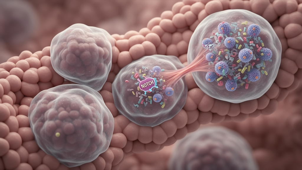
Colorectal cancer diagnosed before age 50, termed early-onset colorectal cancer (EOCRC), is associated with a cellular ecosystem that differs from that of average-onset disease, according to a single-cell transcriptomic analysis of 473,939 cells from 174 patients. The study found that younger-onset tumors were enriched for malignant epithelial states linked to inflammation and proliferation, while their vascular and fibroblast compartments were also remodeled toward proliferative and inflammatory programs. These coordinated differences extended beyond altered chromosome copy number and included selective changes in signaling networks involving NOTCH-associated mediators, although the results identify a candidate biological framework rather than a clinically validated mechanism or treatment target.
The researchers integrated seven publicly available single-cell RNA-sequencing cohorts and, after quality control and Harmony-based batch correction, identified nine major cellular lineages: epithelial cells, endothelial cells, fibroblasts, pericytes, myeloid cells, T cells, B cells, plasma cells and mast cells. The atlas included 39 patients with EOCRC and 135 with average-onset colorectal cancer (AOCRC). Marker genes supported the annotation of each lineage, while quantitative tests and uniform manifold approximation and projection visualizations assessed whether batch correction had removed technical differences without erasing biological variation.
EOCRC tumors contained a higher proportion of epithelial cells and lower fractions of fibroblasts and endothelial cells overall. Immune compartments, by contrast, were comparatively similar between the age groups, indicating that the strongest compositional contrast centered on tumor epithelium and supporting stroma rather than on a wholesale difference in immune representation. Within the epithelial compartment, chromosome-level copy-number variation inference separated 52,791 malignant cells from 17,843 CNV-normal epithelial cells across 166 patients.
Six measures were used to quantify genomic instability: mean CNV fraction, percentage of genome altered, weighted genome instability index, numbers of chromosome arms gained and lost, and total aneuploidy score. AOCRC had nominally higher values for three measures, but the age difference disappeared after adjustment for clinical variables. Microsatellite-instability status, rather than age group, was the dominant correlate of CNV burden, with microsatellite-stable tumors showing higher burden than microsatellite-instability-high tumors in the multivariate model.
No individual chromosome-arm difference remained significant after multiple-testing correction, and sensitivity analyses stratified by MSI status, stage and tumor sidedness did not support a robust age effect. The authors therefore concluded that the aggressive epithelial states seen in EOCRC could not be explained simply by greater chromosomal instability. High-resolution reclustering of the malignant cells resolved nine transcriptional states: Stem-like, UGT-high, Goblet/Secretory, Metastasis-associated, OXPHOS-high, Cycling (S/DNA damage response), Cycling (G2/M), Inflammation-associated and Neuroendocrine-like.
The proliferative states expressed cell-cycle genes including MKI67, TOP2A and CCNB1, while metastatic cells expressed MMP7 and S100A2. Patient-level analyses adjusted for stage, MSI status, sidedness and sex found that EOCRC tumors were significantly enriched for Inflammation-associated and Cycling (S/DNA damage response) cells. AOCRC tumors instead contained relatively more OXPHOS-high and Goblet/Secretory states, corresponding to more differentiated or metabolically quiescent programs.
Leave-one-cohort-out analysis showed that the enrichment of the two younger-onset states remained significant after removal of each individual dataset in six of seven iterations, with covariate-adjusted odds ratios remaining directionally consistent. The result points to a shift in the distribution of malignant cell states rather than the emergence of a completely separate lineage. Trajectory analysis placed Stem-like cells at the origin of several inferred differentiation paths and revealed an ordered progression through inflammatory, proliferative, metastatic, secretory and neuroendocrine-like states.
EOCRC cells appeared more often at intermediate pseudotime positions in the pooled cell-level analysis, whereas AOCRC cells were more represented later in the trajectory. That apparent difference did not persist when pseudotime was summarized for each patient and compared between groups, which yielded a Wilcoxon P value of 0.725. The findings support multiple possible routes of malignant epithelial differentiation but do not show that age itself causes a reproducible shift in developmental timing.
The distinction is important because pseudotime is an inferred ordering of transcriptional states, not a direct measurement of how individual cancer cells evolved. The stromal analysis demonstrated that the younger-onset phenotype was not confined to malignant cells. EOCRC tumors had significantly higher proportions of proliferating or cycling endothelial cells and capillary endothelial cells, whereas AOCRC tumors had more arterial endothelial cells.
The proliferating endothelial population expressed MKI67, TOP2A and CCNB2, while Tip endothelial cells expressed angiogenesis-associated genes including PGF, ESM1 and ANGPT2. Fibroblast and smooth-muscle compartments were similarly heterogeneous, including inflammatory, myofibroblast-like and proliferative cancer-associated fibroblast states. A tendency toward greater representation of WNT5A-positive myofibroblast-like CAFs in EOCRC did not meet the threshold for statistical significance.
Together, these observations indicate a vascular and stromal environment in which proliferative and inflammatory programs are more prominently represented in younger-onset tumors. CellChat and NicheNet analyses were used to infer potentially altered communication among malignant epithelial, endothelial and fibroblast populations. Contrary to a model in which younger-onset tumors would have uniformly stronger signaling, AOCRC had a greater total number of inferred interactions and higher aggregate interaction strength.
EOCRC instead showed selective remodeling of particular epithelial–stromal routes. NOTCH was among the highest-ranked pathways in both age groups, and donor-balanced and leave-one-cohort-out analyses produced comparable normalized information flow. The data therefore do not support the claim that NOTCH signaling as a whole is elevated specifically in EOCRC.
Instead, the results point to age-associated differences in selected communications, with NOTCH-associated ligands such as DLL4 and JAG1 emerging as the most donor-consistent stromal candidates linked to a younger-onset malignant-cell transcriptional program. Candidate stromal signals included DLL1, DLL4, JAG1, FGF2, SERPINE2, PSEN1, SEMA6B, NEO1, COL4A1, HMGB1 and several other mediators spanning NOTCH, growth-factor, extracellular-matrix, axon-guidance and inflammatory pathways. No single fibroblast or endothelial subset accounted for the entire inferred network: WNT5A-positive myofibroblast-like CAFs, IL6-positive inflammatory CAFs, MKI67-positive proliferative CAFs, CCL2-positive inflammatory CAFs, proliferating endothelial cells, capillary endothelial cells and arterial endothelial cells contributed different combinations of candidate signals.
The 33 prioritized target genes were enriched for NOTCH signaling, epithelial and fibroblast proliferation, epithelial-to-mesenchymal transition and cell-cycle regulation. DLL4 and JAG1 showed positive, statistically significant correlations with the target program across the full cohort and every leave-one-cohort-out fold, whereas DLL1 and FGF2 were not consistently correlated. These are computational regulatory predictions based on transcript abundance and prior biological knowledge; they do not demonstrate direct ligand–receptor engagement or establish that a particular signal causes proliferation, invasion or survival.
Clinical relevance was assessed by ranking the 33 target genes according to concordant gene-by-age interactions in two independent colorectal cancer survival cohorts, TCGA-COADREAD with 620 patients and GSE39582 with 585 patients. A five-gene panel comprising HEY1, ZEB1, TWIST1, VIM and FN1 was retained. A one-standard-deviation increase in the panel score was associated with poorer overall survival in EOCRC in both cohorts, with a hazard ratio of 3.43 and a 95% confidence interval of 1.44–8.14 in TCGA-COADREAD, and a hazard ratio of 2.09 with a 95% confidence interval of 1.31–3.34 in GSE39582.
No significant association was detected in AOCRC, and the interaction between panel expression and age group was significant in both datasets. Nevertheless, patient-level panel expression did not differ between the age groups, and the association arose from the gene-by-age interaction rather than a simple increase in the panel among younger patients. Because the genes were selected and evaluated in the same two cohorts, the panel remains a candidate signature requiring independent prospective validation.
CellOracle simulations added a computational link between the NOTCH effector HEY1 and the proliferative malignant state enriched in EOCRC. Setting HEY1 expression to zero or elevating it in an inferred gene-regulatory network produced directional shifts in malignant epithelial states, with simulated overexpression moving the Cycling (S/DNA damage response) population toward the proliferative end of a predefined axis. The knockout effect was comparatively modest, and the analysis did not experimentally manipulate HEY1 in cells or animals.
It therefore cannot establish that HEY1 is required to maintain the younger-onset phenotype, although it is consistent with the hypothesis that increased HEY1 activity may reinforce a proliferative program. The study also has important limitations: it combines public datasets with potential differences in platforms, patient selection and clinical annotation; includes only 39 EOCRC patients; and depends on transcript-based inference that requires protein-level and functional confirmation. Spatial localization, organoid–CAF–endothelial co-culture systems, targeted DLL/JAG–NOTCH perturbations and independent prospective cohorts will be needed to determine whether the inferred network has functional or clinical significance.
The strongest conclusion is that EOCRC is characterized not by one universal genomic lesion or isolated tumor-cell state, but by coordinated epithelial, vascular, fibroblast and signaling remodeling that merits experimental testing.
Subject of Research: Single-cell characterization of cellular and signaling differences in early-onset colorectal cancer
Article Title: Multi-lineage reprogramming and NOTCH-associated signaling remodeling characterize the aggressive biology of early-onset colorectal cancer
Article References: Shi, H., Qiao, L., Su, Q., Gao, H., & Cao, Y. (2026). Multi-lineage reprogramming and NOTCH-associated signaling remodeling characterize the aggressive biology of early-onset colorectal cancer. Frontiers in Oncology, 16, Article 1962237. https://doi.org/10.3389/fonc.2026.1962237
Image Credits: AI Generated
DOI: 10.3389/fonc.2026.1962237
Keywords: early-onset colorectal cancer, single-cell RNA sequencing, tumor microenvironment, NOTCH signaling, malignant epithelial states, cancer-associated fibroblasts, endothelial remodeling, copy-number variation, prognostic biomarkers, computational inference, Multi-lineage, reprogramming