PRKAR2A, which encodes the type IIα regulatory subunit of protein kinase A (PKA), is a candidate spatial router of cyclic-AMP signalling in triple-negative breast cancer (TNBC), but current evidence does not establish that it drives metastasis or qualifies it as a clinical biomarker or therapeutic target. A critical review published in Molecular Biology Reports found that a targeted literature search identified no eligible study demonstrating a causal link between PRKAR2A and a TNBC metastasis endpoint. Instead, the available findings place the gene in four limited association or detection contexts, each compatible with several competing biological models.
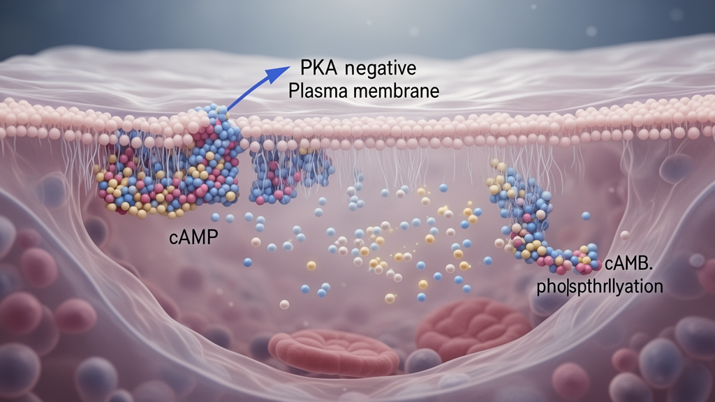
PRKAR2A is notable because its protein product, RIIα, has a dual role in PKA regulation. Within the inactive PKA holoenzyme, RIIα restrains the catalytic subunits. It can also recruit type II PKA to A-kinase anchoring proteins, or AKAPs, which concentrate signalling complexes in defined cellular microdomains. The same subunit can therefore suppress local kinase activity while determining where that activity is positioned. The review proposes that RIIα may act as a driver, a compensatory brake, a localisation determinant or a passenger state marker. Existing evidence cannot discriminate among these models, and the metastasis-specific role of PRKAR2A remains unresolved.
One line of evidence came from an analysis comparing pathological complete response with residual disease in TNBC. PRKAR2A messenger RNA differed near the threshold for statistical significance, but the signal did not survive into the final consensus predictor. This observation suggests a possible association with treatment response or tumour state, yet it does not demonstrate that PRKAR2A causes response, persistence or metastatic progression. A separate experimental context found increased PRKAR2A expression in a lipid-adapted TNBC heterospheroid state. Because the investigators did not perturb the gene specifically, the result cannot establish whether PRKAR2A helps cells adapt to the metabolic environment or merely changes alongside other state-associated transcriptional changes.
RIIα was also detected in fractions containing mitochondria–endoplasmic-reticulum contact sites, where local PKA activity was associated with control of calcium transfer and apoptosis. The experiment linked mitochondrial–endoplasmic-reticulum signalling to a defined biological output, but it did not isolate RIIα as the responsible regulatory component. A fourth line of evidence placed PRKAR2A within a multigene prognostic signature. That inclusion indicates a possible prognostic association, although the gene lacked independent and cell-resolved validation in the evidence reviewed. Together, these observations identify RIIα as a plausible participant in spatially organised cAMP–PKA signalling, rather than a demonstrated metastasis regulator.
Related work involving AKAP2 provides a stronger mechanistic clue but not direct proof for PRKAR2A. Depleting AKAP2 altered focal-adhesion signalling, invasion, primary-tumour growth and lung signal. However, the experiments did not establish which PKA regulatory subunit mediated those effects and did not show a burden-independent role in metastasis. AKAP2 can anchor several signalling systems, so its depletion cannot be treated as a PRKAR2A-specific perturbation. This distinction is central to the review’s conclusion: evidence that an anchoring protein influences tumour behaviour is not equivalent to evidence that PRKAR2A routes cAMP–PKA signals in metastatic cells.
Testing the candidate hypothesis will require experiments designed to separate function, localisation and correlation. The review calls for inducible PRKAR2A depletion rather than reliance on constitutive knockout effects, together with rescue experiments performed at near-endogenous levels and, ideally, with spatial domain specificity. Compartment-targeted PKA reporters would help determine whether PRKAR2A changes where cAMP-dependent phosphorylation occurs. Endogenous mapping of PRKAR2A-containing complexes could establish which AKAPs and regulatory components accompany RIIα in tumour cells. Stage-resolved metastasis assays would then need to test whether manipulating PRKAR2A alters dissemination, survival or outgrowth independently of effects on primary-tumour growth.
Until those decisive tests are completed, the implications should remain narrow. PRKAR2A may help organise the subcellular distribution of PKA, and its expression may mark particular tumour states or contribute to a larger prognostic pattern. The current evidence does not show that manipulating PRKAR2A would prevent TNBC metastasis, improve treatment response or provide a clinically useful biomarker. The review therefore positions RIIα as a candidate spatial signal router and a framework for future experiments, not as a validated target. Its most important unresolved question is whether this dual-function regulatory subunit actively directs metastasis-relevant signalling in the correct subcellular compartment.
Subject of Research: The candidate role of PRKAR2A-encoded RIIα in spatial cAMP–PKA signalling associated with triple-negative breast cancer metastasis.
Article Title: PRKAR2A/RIIα as a candidate spatial cAMP–PKA signal router in triple-negative breast cancer metastasis: evidence boundaries, competing models and decisive experiments
Article References: Muzaffar, T., & Saha, B. (2026). PRKAR2A/RIIα as a candidate spatial cAMP–PKA signal router in triple-negative breast cancer metastasis: evidence boundaries, competing models and decisive experiments. Molecular Biology Reports, 53(1), Article 1768. https://doi.org/10.1007/s11033-026-12975-5
Image Credits: AI Generated
DOI: 10.1007/s11033-026-12975-5
Keywords: PRKAR2A, RIIα, triple-negative breast cancer, cAMP signalling, protein kinase A, cell migration, AKAP2, metastasis, tumour metabolism, critical review, candidate, spatial