In a remarkable stride toward unifying the realms of physics and biology, researchers at the University of California San Diego have published a pivotal review on Raman microscopy, elucidating its profound advances and burgeoning applications within the life sciences. This comprehensive analysis aims to dismantle the barriers between the physicists who engineer these sophisticated instruments and the biologists eager to leverage their capabilities, closing a critical gap that has long hindered interdisciplinary collaboration.
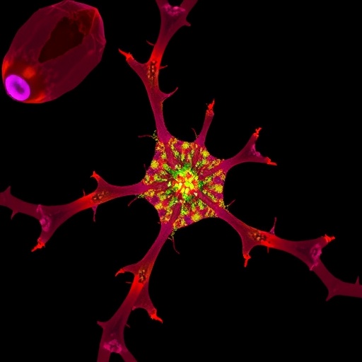
Raman microscopy stands out as a formidable label-free imaging technique capitalizing on the interaction between light and molecular vibrations to generate chemical images of biological specimens. Unlike traditional fluorescent methods, which rely heavily on stains or dyes, Raman imaging harnesses the intrinsic vibrational fingerprints of molecules such as proteins, lipids, DNA, and metabolites. These distinct molecular signatures emerge from the unique frequency shifts in scattered light, offering unparalleled insight into cellular composition and dynamic chemical changes in real time.
Published in the journal Photonix Life, the review is a collaborative work headed by Lingyan Shi, a tenured associate professor of bioengineering at UC San Diego and a visionary in metabolic imaging through platforms such as DO-SRS and SuMMIT-SRS. The article synthesizes a decade’s worth of breakthroughs, providing a resource that bridges technological innovation with practical biology, fulfilling a need long felt by researchers navigating this interdisciplinary space.
Shi emphasized that despite tremendous technical progress, a disconnect remains between what Raman microscopy hardware can accomplish and biologists’ awareness of these possibilities. The review responds directly to this issue, systematically laying out the strengths and limitations of current Raman techniques in a manner accessible to non-specialists. This effort not only democratizes the knowledge but also catalyzes more informed deployment of Raman methods in biological investigations.
One of the review’s central contributions is the establishment of a structured analytic framework to compare Raman imaging technologies across four critical performance metrics: signal sensitivity, imaging speed, three-dimensional volumetric imaging ability, and spatial resolution. This framework spans both spontaneous and coherent Raman modalities, allowing scientists to discern the trade-offs inherent in different approaches and select tools precisely tuned to their experimental questions.
Raman microscopy’s versatility shines as it scales from subcellular observation to clinical application. The review spotlights compelling examples where Raman imaging tracks metabolic shifts during aging and neurodegeneration by employing heavy water as a bio-orthogonal tracer, revealing cellular turnover with exquisite specificity. In neurodegenerative disease research, label-free detection of amyloid plaques addresses the pressing challenge of visualizing pathological aggregates in Alzheimer’s brain tissue without invasive dyes.
Another paradigm-shifting application detailed is the use of AI-powered virtual staining that condenses conventional histological preparation times from over 36 hours to a mere 3 minutes, facilitating near-instantaneous diagnostic tissue assessment. Similarly, real-time imaging of drug diffusion into tissues opens doors for more precise pharmacokinetic studies and could revolutionize personalized medicine approaches.
From a technical perspective, the review chronicles several groundbreaking advances pushing the very limits of Raman microscopy. Noteworthy are tip-enhanced Raman methods achieving spatial resolutions as fine as 2.5 nanometers—this nanoscale precision suffices to differentiate individual protein complexes on cellular membranes, a milestone once thought unattainable. Concurrently, advancements in quantum-enhanced detection using squeezed light surpass classical noise floors, amplifying signal strength by over 50% and enabling faster, more sensitive imaging of live cells without photodamage.
The integration of computational innovations further refines imaging resolution. The A-PoD super-resolution algorithm, developed in Shi’s laboratory, exemplifies how software-driven post-processing can breach the 59-nanometer barrier, rendering molecular details with unprecedented clarity. This synergy between hardware and point computational techniques signifies a new era of accessible super-resolution imaging.
Looking forward, the review heralds several emerging frontiers poised to redefine biomedical imaging. Miniaturized fiber-optic Raman probes boasting over 98% accuracy in cancer diagnostics during endoscopy promise transformative clinical adoption, bringing the power of molecular imaging directly into patient care settings. Quantum coherent effects, enabling selective amplification of target molecular signals, expand the repertoire of measurable biological processes with exceptional specificity.
Furthermore, ultrafast time-resolved Raman spectroscopy facilitates observation of molecular dynamics on femtosecond timescales, capturing biochemical events previously obscured by temporal constraints. The fusion of Raman microscopy with mass spectrometry and clinical MRI in multimodal platforms presents a holistic approach to tissue characterization, integrating chemical, structural, and metabolic data streams into comprehensive diagnostic landscapes.
Erick Alvarado, a doctoral candidate and primary author of the review, reflects on the field’s trajectory: “The technology is mature enough to tackle real-world biological and clinical challenges, but the real breakthrough lies in fostering dialogue between physicists and biologists. Our review aims to build this common language, accelerating the translation of these powerful tools from lab benches to bedside applications.”
As Raman microscopy moves decisively beyond experimental confines toward widespread adoption in clinical and industrial spheres, the fusion of quantum-enhanced imaging, AI-assisted diagnostics, and compact probe design is set to establish it as a cornerstone in precision medicine. This confluence promises unprecedented access to real-time, single-molecule, and multidimensional molecular insights, propelling life sciences into a transformative new era.
The extensive review by UC San Diego’s bioengineering team not only serves as an indispensable technical guide but also as a clarion call for a collaborative vision—one where disparate disciplines unite to fully harness the promise of label-free chemical imaging. The future of health and biology stands to be profoundly reshaped by the innovations chronicled in this landmark synthesis.
Subject of Research: Not applicable
Article Title: Raman microscopy unmixed: Technical advances, bio-orthogonal tags, and applications in life sciences
Web References: http://dx.doi.org/10.3724/PXLIFE.2025-0016
References: See article via doi link
Keywords: Raman microscopy, biomedical imaging, label-free imaging, metabolic imaging, quantum-enhanced imaging, super-resolution, AI, precision medicine, bio-orthogonal tags, molecular vibrations, coherent Raman, spontaneous Raman
Tags: advances in Raman imaging technologybioengineering applications of Raman microscopychemical imaging of biological specimensDO-SRS and SuMMIT-SRS imaging platformsinterdisciplinary collaboration in biophysicslabel-free molecular imaging techniquesmetabolic imaging advancementsmolecular vibrations in microscopyprotein and lipid imaging with RamanRaman microscopy in biological researchRaman spectroscopy for DNA and metabolitesreal-time cellular composition analysis