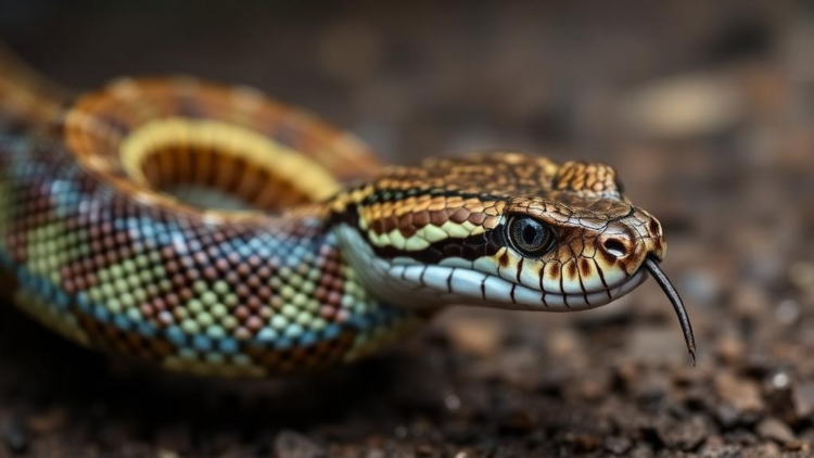
Boid inclusion body disease, or BIBD, has haunted captive boa constrictors since the late 1970s, quietly spreading through private collections, breeding colonies, and zoos around the world. The disease is caused by reptarenaviruses, a group of genetically highly divergent arenaviruses that were only identified as the causative agents in the early 2010s. Its diagnostic hallmark is the presence of intracytoplasmic inclusion bodies, dense structures inside cells that are composed primarily of the roughly 68 kilodalton viral nucleoprotein. For decades, veterinarians have observed that snakes with BIBD frequently suffer from bacterial, fungal, and protozoal co-infections, and this pattern gave rise to a widely accepted hypothesis: that the virus somehow suppresses the immune system of its host, leaving the animal vulnerable to opportunistic pathogens. Yet, despite how often this idea has been repeated, an immunosuppressive effect had never actually been confirmed for reptarenaviruses in their snake hosts.
A new study published in iScience by Eva Dervas of the University of Zurich and colleagues set out to test that long-standing assumption directly. The researchers assembled a remarkable cohort of 133 captive boa constrictors drawn from breeding colonies, private collections, and a reptile shelter in Switzerland and Germany. Using blood smears, the gold standard diagnostic method, they identified 49 animals with BIBD based on the presence of inclusion bodies in peripheral blood cells. A multiplex reverse transcription PCR confirmed reptarenavirus infection in every one of those animals and, crucially, identified three additional snakes that carried viral RNA without any detectable inclusion bodies, making them asymptomatic carriers. The remaining 83 animals tested negative for both disease and virus. The cohort spanned ages from three months to 25 years, allowing the team to disentangle the effects of disease from those of normal aging and sex.
The researchers hypothesized that BIBD compromises both innate and adaptive immune functions, and that these impairments would be reflected in altered blood parameters, impaired antibody responses, and elevated stress hormones. To test this, they performed an unusually comprehensive battery of assays on blood and plasma samples. These included hematological and biochemical analyses, corticosterone measurements, a phagocytosis assay using fluorescent beads, lysozyme quantification, hemagglutination and hemolysis tests for natural antibodies and complement, a leukocyte blast transformation test for T cell function, and enzyme-linked immunosorbent assays for virus-specific immunoglobulins. Few studies of reptilian immunology have attempted such breadth, and the effort reflects how little is still known about how snake immune systems work.
The first striking finding concerned phagocytosis, the process by which immune cells engulf and destroy pathogens. In boa constrictors, two related cell types perform this function in the blood: monocytes and azurophils, the latter being a cell type unique to reptiles whose exact origin and function remain poorly understood. Both express Iba1, a marker of the monocyte and macrophage lineage. When the researchers incubated white blood cells with fluorescent beads, they found that BIBD-positive snakes had a significantly lower percentage of Iba1-positive cells performing phagocytosis, with a mean of 52.2 percent compared with 64.36 percent in uninfected animals. BIBD-positive snakes also had significantly fewer circulating monocytes, with a geometric mean percentage of 1.62 percent versus 2.4 percent in disease-free animals. These deficits held up after statistical correction for age, sex, and clustering by breeder, and they were also evident when animals were grouped simply by infection status rather than by disease.
This impairment of phagocytic cells is particularly intriguing when viewed against what is known about mammalian arenavirus infections. In humans infected with Lassa virus or lymphocytic choriomeningitis virus, infection and inhibition of macrophages and dendritic cells are well-documented features of disease. Lassa virus infection reduces the phagocytic activity of dendritic cells and macrophages, an effect proposed to stem from a failure of monocyte maturation. More recent work on New World arenaviruses such as Junín and Machupo viruses has uncovered mechanistic links between viral entry and phagocytosis, identifying host restriction factors like TRIM2 and SIRPA that jointly regulate both processes. Although the receptors used by reptarenaviruses to enter snake cells are still unknown, the authors suggest that the reduced phagocytic activity they observed could represent a direct consequence of the virus infecting these very cells. Notably, their earlier work showed that the inclusion body-laden cells of the bone marrow, including hematopoietic precursors, are infected in BIBD, and that monocytes and azurophils themselves carry viral inclusion bodies.
Not every component of the immune system showed damage. Lysozyme concentrations, natural antibody and complement titers measured by hemagglutination and hemolysis, and the leukocyte blast transformation response to phytohemagglutinin stimulation all remained unchanged between diseased and healthy animals. Stress markers told a similar story of stability: plasma corticosterone, glucose, and the heterophil-to-lymphocyte ratio did not differ significantly between groups, although the authors caution that variable sampling conditions, with some animals handled briefly at home and others transported before sampling, likely introduced considerable interindividual noise. The findings suggest that BIBD is not a blanket collapse of immunity but a targeted disruption, with the phagocytic arm of innate immunity bearing the brunt of the impact.
The adaptive side of the immune response revealed a more nuanced picture. Snakes with BIBD had significantly lower total plasma immunoglobulin concentrations, with a geometric mean of 11.23 percent compared with 16.97 percent in uninfected animals. Yet when the researchers measured antibodies specific to the reptarenavirus University of Giessen virus 1, the diseased snakes showed higher levels of both virus-specific IgM and IgY than uninfected animals. Using established cutoff values, 77.1 percent of BIBD-positive snakes tested positive for anti-UGV-1 antibodies, with IgM detected more frequently than IgY, an unexpected result given that IgY, the functional equivalent of mammalian IgG, is generally considered the dominant antibody class in reptiles. Interestingly, 36.7 percent of uninfected snakes also carried anti-UGV-1 antibodies, hinting at either past infections that were cleared or cross-reactive responses to undetected viruses. The authors propose several explanations for the paradox of low total immunoglobulin alongside high virus-specific titers, including general dampening of antibody production by the virus or the sequestration of antibodies into circulating immune complexes, a phenomenon documented in persistent lymphocytic choriomeningitis virus infections of mice.
Beyond immunity, the study uncovered a metabolic signature of disease. BIBD-positive snakes had significantly lower blood cholesterol than their uninfected counterparts, and the same held true when animals were grouped by infection status. The authors note that hypocholesterolemia in mammals is associated with several chronic infections, including HIV and pulmonary tuberculosis, where lower cholesterol correlates with disease severity and even mortality. Because the snakes came from multiple collections with varying husbandry and showed no significant weight loss, diet is an unlikely explanation. Instead, the researchers point to the liver: inclusion bodies form in the majority of hepatocytes in BIBD, and their previous work demonstrated that the viral nucleoprotein accumulates within the mitochondria of infected cells. Given the central role of hepatocyte mitochondria in lipid metabolism, viral-induced mitochondrial dysfunction could plausibly underlie the observed drop in cholesterol, a hypothesis the authors hope future studies will test directly.
The study also carefully separated the effects of disease from those of ordinary biological variation. Regardless of BIBD status, heterophil and azurophil percentages and hematocrit rose with age while lymphocyte percentages fell, mirroring the shift from lymphoid-dominated to myeloid-dominated hematopoiesis seen in mammals. Aspartate transaminase levels correlated with male sex rather than disease, echoing the sex differences seen in human reference ranges. These controls strengthen the conclusion that the immune and metabolic changes tied to BIBD are genuine disease effects rather than artifacts of demography. The authors acknowledge limitations, including the cross-sectional design, the small number of carrier animals, and the rarity of clinically affected individuals in the cohort, and they call for longitudinal and experimental infection studies to determine whether reduced phagocytosis precedes overt disease and could serve as an early warning sign. For now, the work delivers a clear verdict on a decades-old assumption: BIBD does alter immune function in boa constrictors, but selectively, impairing phagocytic innate defenses and reshaping antibody dynamics in ways that may finally explain why these snakes so often fall to secondary infections.
Subject of Research: Immune function alterations in boa constrictors with boid inclusion body disease caused by reptarenavirus infection
Article Title: Boid inclusion body disease in Boa constrictor : Unraveling the myth of immunosuppression
Article References: Dervas, E., Czirják, G. Á., Michalopoulou, E., Liesegang, A., Novacco, M., Schwarzenberger, F., Hetzel, U., Hepojoki, J., Baggio, F., & Kipar, A. (2026). Boid inclusion body disease in Boa constrictor: Unraveling the myth of immunosuppression. iScience, 29(10), Article 117669. https://doi.org/10.1016/j.isci.2026.117669
Image Credits: AI Generated
DOI: 10.1016/j.isci.2026.117669
Keywords: boid inclusion body disease, reptarenavirus, Boa constrictor, immunosuppression, phagocytosis, innate immunity, adaptive immunity, azurophils, monocytes, cholesterol, IgY antibodies, reptile immunology
Cite Scienmag News
APA MLA Chicago
Copy citation Download RIS
Tags: adaptive immunityazurophilsBIBD diagnosis and symptomsBoa constrictorboa constrictor diseaseboid inclusion body diseasecaptive snake health studiescholesterolIgY antibodiesimmune system effects in reptilesimmunosuppressioninnate immunitymonocytesnovel insights into snake virus impactphagocytosisreptarenavirusreptarenavirus immune suppressionreptile co-infectionsreptile disease transmissionreptile immunologyreptile infectious diseasesreptile virology researchSnake virusviral nucleoprotein in snakes