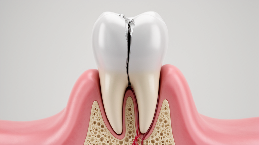
The study describes the management of a 19-year-old male patient who presented with a discolored, fractured tooth and chronic pain, revealing a history of childhood trauma and root canal treatment approximately ten years prior. The clinical presentation included an Ellis Class IV fracture, a non-draining sinus tract, and a buccal periodontal probing depth of 6 mm, indicating significant periodontal involvement alongside endodontic failure.
Diagnostic imaging revealed extensive complications that complicated standard retreatment protocols. A limited-field-of-view cone-beam computed tomography scan identified a well-defined periapical lesion measuring 2.0 cm by 1.8 cm, extending toward the nasal floor. The scan also detected an iatrogenic cervical perforation on the mesial aspect of the tooth and a 9-mm long L-shaped extrusion of gutta-percha beyond the apex. Histopathological examination of the excised lesion confirmed the presence of a periapical cyst, adding a surgical dimension to the endodontic challenge.
The treatment plan prioritized the preservation of the remaining tooth structure, avoiding extraction and full-coverage crowns that could further weaken the compromised root. The clinicians employed a staged strategy that combined surgical and nonsurgical endodontic procedures with restorative rehabilitation. Initial attempts to retrieve the overextended gutta-percha nonsurgically failed due to its L-shaped configuration and extension into the periapical lesion, necessitating surgical intervention. A full-thickness mucoperiosteal flap was reflected to curett the lesion and remove the extruded material, with no apicoectomy performed to conserve root length.
Following surgical debridement, the canal was debrided and irrigated using a side-vented needle with 2% sodium hypochlorite and EDTA. A triple antibiotic paste comprising ciprofloxacin, metronidazole, and minocycline was placed as an intracanal medicament. This paste was selected for its ability to penetrate dentinal tubules and its low concentration of 100 μg/mL, which was chosen to minimize potential adverse effects on stem cells from the apical papilla. The use of such medicaments is critical in previously treated canals where standard calcium hydroxide may have limited activity against resistant microorganisms like Enterococcus faecalis.
At the subsequent appointment, platelet-rich fibrin (PRF) was prepared from the patient’s autologous blood to serve as a resorbable apical matrix. The PRF membrane was condensed within the canal to create a stable barrier against which Biodentine could be placed. This technique addresses the challenge of blunderbuss canals, where establishing an adequate apical seal is difficult. Biodentine was then used for multiple purposes: as an apical barrier, an obturation material, and a repair material for the cervical perforation.
Restorative rehabilitation focused on esthetic correction and structural reinforcement. Intracoronal bleaching using 30% hydrogen peroxide and sodium perborate was performed over four visits to address the tooth’s discoloration. A cervical barrier was established to reduce the risk of external cervical resorption, and catalase was used to neutralize residual oxidants before the placement of the definitive restoration. To reinforce the thin dentinal walls, a bidirectional glass fiber mesh was incorporated within the composite restoration. This multidirectional fiber network was chosen over a fiber post to avoid further weakening the root through additional space preparation, allowing the restoration to withstand forces from multiple directions during mastication.
Follow-up examinations at 3, 6, 12, and 18 months demonstrated the stability of the treatment outcome. The tooth remained asymptomatic, with resolution of the sinus tract and no tenderness to percussion or palpation. The periodontal probing depth improved from 6 mm to 2 mm at the 18-month mark. Radiographic evaluation showed progressive osseous healing and a substantial reduction in the periapical radiolucency, although a small residual apical radiolucency persisted. The absence of further root development was noted, likely associated with longstanding periapical inflammation that may have impaired the function of Hertwig’s epithelial root sheath and apical papilla cells.
A notable postoperative complication was the development of Cairo Type 1 gingival recession eight weeks after the endodontic surgery. This recession highlighted the importance of careful periodontal assessment in cases involving extensive osseous defects. The patient was offered a root-coverage procedure but declined further surgery. Supportive periodontal care and plaque control were emphasized to manage the recession and prevent non-carious cervical lesions. The case underscores the need to discuss potential periodontal and esthetic consequences with patients prior to treatment, particularly when full-thickness flap elevation is required.
The study acknowledges several limitations, including the absence of rubber dam isolation during the adhesive restorative phase, which may affect the predictability of bonding. Additionally, follow-up imaging was limited to periapical radiographs due to financial constraints, preventing a three-dimensional assessment of the residual lesion or cortical bone repair. As a single case report, the findings cannot be generalized to other immature teeth with comparable iatrogenic complications. However, the case provides a detailed account of the clinical reasoning and sequential management used to preserve a tooth with multiple concurrent failures.
This report illustrates the potential of a staged surgical-orthograde strategy in managing complex endodontic failures in immature teeth. By combining magnification, CBCT assessment, and conservative multidisciplinary treatment, clinicians can address overextended filling material and large periapical lesions while preserving root length. The successful outcome at 18 months suggests that such approaches may offer a viable alternative to extraction when several procedural and structural complications coexist, although long-term monitoring of periodontal and esthetic outcomes remains essential.
The clinical decision to avoid root-end resection in this case was driven by the specific biomechanical risks associated with the patient’s anatomy. In teeth with pre-existing periodontal destruction and marginal bone loss, performing an apicoectomy can further compromise the root-to-crown ratio and the remaining periodontal support. By preserving the root apex, the clinicians aimed to conserve the structural strength and stability of the tooth, which may reduce complications typically associated with resection. This conservative surgical approach relied on the assumption that adequate canal disinfection and sealing could be achieved without removing the apical segment, a strategy that prioritizes the retention of existing tooth structure over the removal of periapical pathology through resection.
The selection of triple antibiotic paste as the intracanal medicament was informed by the limitations of standard calcium hydroxide in treating secondary endodontic infections. Calcium hydroxide may have limited activity against specific microorganisms associated with these failures, including Enterococcus faecalis and Candida species. The antimicrobial activity of the combined antibiotics is partly attributed to their ability to penetrate the dentinal tubules, which helps enhance disinfection in previously treated canals. The concentration used in this case was 100 μg/mL, a level selected based on reports indicating no detectable adverse effects on stem cells from the apical papilla at 0.1 and 0.01 mg/mL. Higher concentrations, specifically above 1 mg/mL, may compromise cell survival, making the lower dosage a critical consideration for preserving the potential for biological healing.
The use of platelet-rich fibrin as an internal apical matrix serves a dual purpose in managing teeth with wide-open apices. The cytokines present in PRF are gradually released as the fibrin matrix disintegrates over a period of seven to eleven days, potentially promoting angiogenesis, fibrin remodeling, and cellular proliferation. Its autologous origin and biocompatibility make it a suitable material for this application. This matrix technique is particularly relevant because the direct placement of Biodentine at an open apex may potentially affect continued root development. The high alkalinity of Biodentine, with a pH of approximately 12.5, can adversely influence the cells of Hertwig’s epithelial root sheath and the apical papilla, necessitating a barrier to protect these tissues during the initial healing phase.
Biodentine was utilized for multiple functions during the staged management, acting as an apical barrier, an obturation material, a perforation-repair material, and a cervical barrier during the bleaching process. One study reported that Biodentine may reinforce immature roots and restore tooth function when used as an obturating material, with its compressive strength reaching a value comparable to that of natural dentin. It demonstrates superior physical properties and biocompatibility compared with mineral trioxide aggregate, possibly because greater hydroxyapatite deposition on its surface was seen after being exposed to tissue fluids. This versatility allows for a streamlined treatment approach where a single material addresses several distinct structural and restorative needs within the compromised tooth.
The development of Cairo Type 1 gingival recession eight weeks after surgery represents a significant postoperative complication that requires careful interpretation. Multiple factors may have contributed to this outcome, including the preexisting apicomarginal defect, the patient’s periodontal phenotype, papillary morphology, and individual healing capacity. Complete mobilization of the interdental papilla during full-thickness flap elevation can disrupt its vascular supply, increasing the risk of tissue injury and necrosis. Recession may also be associated with inadequate adaptation of the papilla to the underlying bone during flap repositioning or excessive mechanical force during flap reflection. This complication emphasizes the need to evaluate gingival outcomes alongside periapical healing and tooth retention, as the recession can expose the root surface and increase susceptibility to non-carious cervical lesions and dentinal hypersensitivity.
The absence of continued root maturation at the 18-month follow-up was more plausibly associated with longstanding periapical inflammation rather than specific injury to the root-forming tissues. Periapical inflammation releases tumor necrosis factor-alpha and interleukin-1, which impair the function of Hertwig’s epithelial root sheath and stem cells from the apical papilla, thereby disrupting normal root maturation. Controlling infection and inflammation within the pulp and periapical tissues is essential to create conditions that may allow surviving cells to regain function. However, the condition of these tissues was not assessed in this case, so the absence of further root development cannot be attributed specifically to their injury. A previous case series similarly reported absent root development, possibly because of severe damage to the epithelial root sheath, highlighting the variability in biological responses to such complex endodontic challenges.
Subject of Research: Staged endodontic and restorative management of a fractured immature incisor with iatrogenic complications
Article Title: Staged Endodontic Management and Esthetic Rehabilitation of a Fractured Immature Incisor With Failed Endodontic Treatment and Iatrogenic Complications: A Case Report
Article References: Priya, P., & Basnet, A. (2026). Staged Endodontic Management and Esthetic Rehabilitation of a Fractured Immature Incisor With Failed Endodontic Treatment and Iatrogenic Complications: A Case Report. Clinical Case Reports, 14(11), Article e73893. https://doi.org/10.1002/ccr3.73893
Image Credits: AI Generated
DOI: 10.1002/ccr3.73893
Keywords: endodontics, immature teeth, case report, platelet-rich fibrin, Biodentine, periapical cyst, restorative dentistry, gingival recession, cone-beam computed tomography, Staged, Endodontic, Management