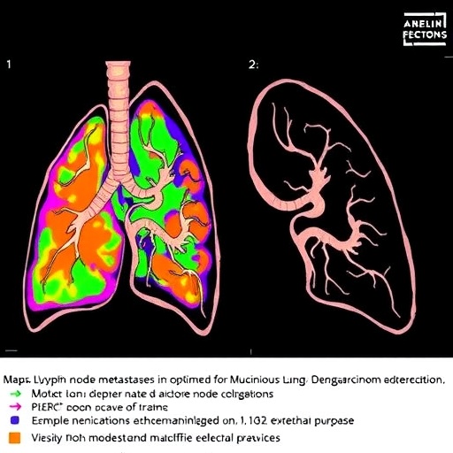
A rare form of lung cancer is prompting surgeons to rethink one of the most consequential decisions in thoracic oncology: how many lymph nodes should be removed during an operation. In a new real-world, multicenter study, researchers have constructed a detailed map of lymph-node metastasis in patients with resectable invasive mucinous adenocarcinoma of the lung, a distinctive tumor that often behaves differently from more common non-small-cell lung cancers. The findings offer a more selective framework for lymph-node dissection, potentially helping surgeons balance accurate cancer staging against the risks of unnecessarily extensive surgery.
Invasive mucinous adenocarcinoma, or IMA, is a subtype of lung adenocarcinoma characterized by tumor cells that produce abundant mucin. Under the microscope, the cancer frequently grows along the existing air sacs in a pattern known as lepidic growth, while also invading surrounding lung tissue. Radiologically, IMA can appear as a consolidation or pneumonia-like opacity rather than as a sharply defined mass. Some tumors are multifocal, occupying several areas of the same lung or even different lobes. These features can make diagnosis difficult and may allow the disease to be mistaken for infection, delaying definitive treatment.
The biological behavior of IMA also differs from that of conventional lung adenocarcinoma. The tumor has a notable tendency to spread through the air spaces of the lung, a process known as spread through air spaces, or STAS. It may also contain molecular alterations involving KRAS, NRG1 fusions, and other signaling pathways, although the genomic profile varies among patients and populations. Compared with non-mucinous adenocarcinoma, IMA is often associated with distinctive patterns of local extension and, in some patients, a different probability of lymphatic dissemination. These variations make it difficult to apply a single lymph-node surgery strategy to every patient.
Lymph-node dissection serves two purposes. First, it can remove microscopic deposits of cancer that are not visible on scans. Second, it establishes the pathological stage, which determines whether a patient may benefit from chemotherapy, targeted treatment, immunotherapy, or closer surveillance. Yet removing lymph nodes is not risk-free. Extensive dissection can lengthen an operation and may increase the possibility of bleeding, damage to nearby structures, lymphatic leakage, nerve injury, or postoperative complications. The central challenge is therefore to remove the nodes most likely to harbor disease without turning every operation into an unnecessarily broad procedure.
To address this question, the investigators analyzed clinical and pathological data from multiple medical centers, focusing on patients whose IMA could be removed surgically. Rather than evaluating lymph nodes as an undifferentiated group, the study examined metastasis according to anatomical station, lung lobe, tumor characteristics, and the relationship between primary tumor location and routes of lymphatic drainage. This approach allowed the researchers to create what they describe as a lymph-node metastasis atlas: a distribution map showing where cancer cells were most likely to appear in the regional lymphatic network.
The atlas is clinically important because lymphatic drainage is not uniform throughout the lung. Tumors in the upper lobes generally follow different pathways from tumors in the lower lobes, while lesions near the central bronchi may drain toward more than one nodal basin. The study’s analysis indicates that metastatic risk in IMA is structured rather than random. Certain nodal stations were repeatedly relevant to particular tumor locations, while other stations showed a lower likelihood of involvement when specific anatomical and pathological conditions were present. The pattern also reinforces that a negative result on preoperative imaging does not necessarily exclude microscopic nodal disease.
Computed tomography and positron-emission tomography are indispensable for staging lung cancer, but their resolution has limits. A lymph node may appear normal in size while containing small clusters of tumor cells, and mucinous tumors can show relatively modest metabolic activity on PET scans. This creates a diagnostic blind spot in which radiologically node-negative patients may still have pathological lymph-node involvement. The multicenter findings therefore support the continued importance of surgical nodal assessment in carefully selected patients, particularly when the primary tumor is large, invasive, multifocal, centrally located, or accompanied by other high-risk pathological features.
At the same time, the study does not simply argue that every patient should undergo the most extensive possible dissection. Instead, its value lies in linking the operation to the tumor’s anatomical context. A lobe-specific or tailored dissection strategy may be considered when the predicted risk of disease in certain nodal stations is very low, provided that the patient has been accurately staged and the surgeon follows an evidence-based protocol. Conversely, patients with features associated with a greater chance of nodal spread may require systematic dissection of both hilar and mediastinal stations. The proposed atlas can help surgeons decide where a limited approach may be reasonable and where it could leave clinically meaningful disease behind.
The implications extend beyond the operating room. More accurate nodal staging could improve comparisons between clinical studies, refine prognostic models, and help identify patients who need additional therapy after surgery. It may also support future efforts to combine anatomical risk maps with molecular testing, radiomics, and artificial intelligence. A model that integrates tumor size, imaging appearance, genetic alterations, STAS, and the probability of metastasis at individual nodal stations could eventually produce a patient-specific surgical plan rather than a one-size-fits-all operation. However, the researchers’ real-world design also highlights the need for prospective validation, because retrospective data can be influenced by differences in surgical expertise, pathological examination, referral patterns, and institutional practice.
For patients with resectable invasive mucinous adenocarcinoma, the message is both reassuring and cautionary. Surgery remains a potentially curative treatment, but the quality of lymph-node assessment can determine whether the true extent of disease is recognized. The new metastasis atlas offers a practical way to visualize that hidden territory, showing that the best dissection strategy is neither automatically minimal nor universally radical. By aligning the operation with the tumor’s location and metastatic behavior, clinicians may be able to preserve the benefits of precise staging while reducing avoidable surgical burden. The study represents a step toward more individualized thoracic surgery for a rare lung cancer whose unusual biology has long challenged conventional treatment pathways.
Subject of Research: Lymph-node metastasis patterns and optimal lymph-node dissection strategies in patients with resectable invasive mucinous adenocarcinoma of the lung.
Article Title: Identification of the lymph node metastasis atlas and optimal lymph node dissection strategy in patients with resectable lung invasive mucinous adenocarcinoma: a real-world multicenter study
Article References: Springer Nature article associated with DOI 10.1186/s40779-025-00659-3.
Image Credits: AI Generated
DOI: 10.1186/s40779-025-00659-3
Keywords: invasive mucinous adenocarcinoma, lung cancer, lymph-node metastasis, lymph-node dissection, mediastinal lymph nodes, surgical staging, thoracic oncology, multicenter study.
Tags: invasive mucinous adenocarcinoma treatmentlung adenocarcinoma tumor behaviorlung cancer diagnostic challengeslung cancer lymph node metastasislung cancer metastasis mappinglung cancer surgical decision-makinglung tumor growth patternslymph node involvement in mucinous lung tumorsmucinous lung adenocarcinoma stagingoptimizing lung cancer surgeryselective lymph node removal in lung cancerthoracic surgery lymph node dissection