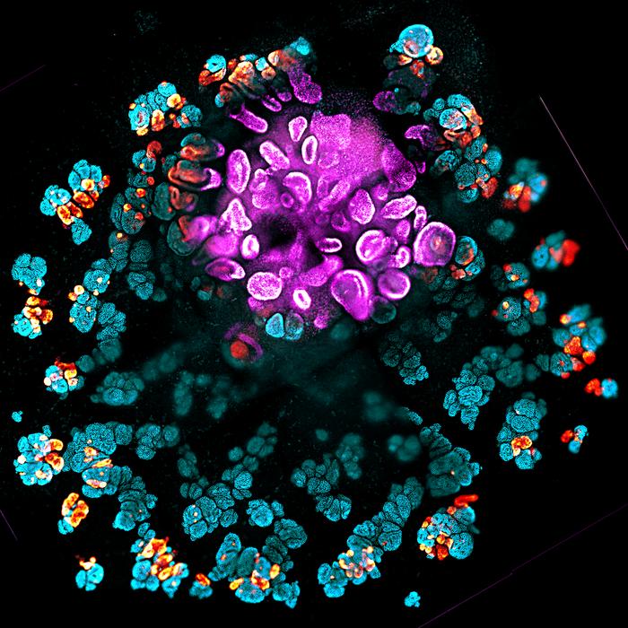
![Kidney organoid [Lindstrom Lab/USC Stem Cell]](https://www.genengnews.com/wp-content/uploads/2026/07/low-res-1.jpeg "Kidney organoid")
University of Southern California (USC) researchers have paired a biological discovery with an engineering feat to create more faithful, reproducible kidney organoid structures, grown from human pluripotent stem cells (hPSCs). By mapping the developing human kidney, the scientists identified a previously unrecognized developmental axis that helps organize the kidney’s nephrons, which are their filtering units. The team then engineered Wnt-secreting “synthetic organizer” cells to recreate aspects of this developmental environment in organoids.
Their advance makes the organoids more reliable models for studying disease and evaluating potential therapies, while supporting long-term efforts to generate transplantable kidney tissue. “It is important that we’re starting to get good reproducibility from organoid models that can lead to robust preclinical models of cell function and disease to benefit patients,” said Nils Lindström, PhD, assistant professor of stem cell biology and regenerative medicine at the Keck School of Medicine of USC. Lindström is co-corresponding author of the team’s published report in Science, titled “Patterning human kidney organoids with synthetic Wnt-secreting organizers.” In their paper, the researchers reported, “Our findings link a spatial organizing geometry in the developing human kidney to controllable engineering in vitro.”
“Stem cell–derived organoids have emerged as systems for modeling organ development and generating complex tissue structures in vitro,” the authors wrote. Over the past decade, organoid work has relied on cells’ ability to self-organize into tissue-like structures, often in response to adding chemicals and proteins that act broadly in the whole organoid. “Although this capacity enables organoids to recapitulate many developmental programs, it limits experimental control over tissue architecture, often producing structures that vary between cultures and are difficult to engineer reproducibly,” the team continued. “Understanding how to impose spatial patterning in organoid systems is therefore an important challenge.”
In embryos, spatial patterning is often organized by localized signalling centers, known as developmental organizers. But how organizing geometry is controlled in the developing kidney, and whether it can be recreated in vitro, hasn’t been known.
For their reported study, the team combined spatial transcriptomics of the developing human kidney with synthetic engineering. “We mapped this organizing geometry in developing human kidneys and tested whether a minimal cue of localized WNT signaling could restore spatial control of nephron patterning in organoids,” they explained.
The project began by making tools to copy developmental signals. Postdoctoral researcher Fokion Glykofrydis, PhD, in the Morsut lab, engineered a “synthetic organizer” cell that secreted a Wnt protein that their spatial transcriptomics and other analyses indicated was involved in spatial patterning during kidney development. Graduate student Connor Fausto from the Lindström lab proposed an experiment to test how this Wnt-secreting cell would affect organoid nephrons.
The experiments revealed that the synthetic organizer enabled two key processes essential for building organs: controlling the identity of cells and influencing the shape of developing structures. The synthetic organizer serves as a localized and targeted source that secretes controllable amounts of specific Wnt proteins within the organoid itself. These are key signals that help shape the developing kidney. This creates a signaling environment much more similar to a naturally developing kidney and gives researchers a way to control where and how kidney structures form.
Co-corresponding author Leonardo Morsut, PhD, associate professor of stem cell biology and regenerative medicine, and biomedical engineering at the Keck School of Medicine and USC Viterbi School of Engineering, said, “With our approach, we are trying to control self-organization, and work with it as opposed to try to completely override it.”
Lindström expected Wnt to trigger nephrons to change their identity into cells capable of forming connections with the urine drainage system. What surprised him was that the nephrons also changed shape and elongated toward the source of the Wnt signal, which doesn’t happen when signals are delivered uniformly to the whole organoid. Compared with the developmental process seen in traditional kidney organoids, this elongation toward the Wnt source is more similar to what happens in a naturally developing kidney.
“A single, localized signal did two things at once. It changed what the cells became and physically pulled the tubules toward the source,” Lindström said. “You would not see that with a uniform chemical bath of signals.” Engineered WNT-secreting cellular organizers introduced into kidney organoids restored organizing geometry, the authors noted, “… biasing distal nephron differentiation and orienting nephron morphogenesis toward the signal source, which demonstrates that developmental signaling geometry can be reconstructed synthetically to control tissue patterning.”
The team identified a previously unrecognized axis, a direction along which the developing kidney organizes itself. Developmental biologists have long known about the nephron’s classic “proximal-distal (PD) axis,” which runs from its blood-filtering end to its urine-drainage end. The new axis is defined instead by how close each part of the nephron sits to the collecting duct, the tube system that drains urine and releases Wnt signals during development. Those signals tell the nephron what shape to take and which way to point.
“The study shows that there’s an undiscovered axis that sets up how a nephron looks and forms,” said Lindström. “It’s not every day that you find something new in human development at that level.”
Most kidney organoids contain only nephrons and lack the collecting duct that supplies this local Wnt signal, so they have no such axis and organize in a radially symmetrical pattern. By mapping how kidney cells respond to Wnt at specific locations in the developing kidney, the team recreated that environment in organoids with the synthetic organizer, producing structures that are both more developmentally faithful and more reproducible.
“Introducing tunable WNT-secreting synthetic organizers (SOs) in organoids restored canonical WNT responses, biased distal nephron differentiation, and oriented nephron morphogenesis toward the WNT source,” the investigators stated in summary. The combined results, they suggested, “… demonstrate that the spatial geometry observed in vivo can be reconstructed synthetically to control early nephron patterning and morphogenesis … Synthetic organizers provide a modular way to restore missing spatial interactions without reconstructing the entire collecting duct lineage, complementing approaches that rebuild collecting duct–to–nephron cellular interactions.”
For Morsut, the synthetic organizer is one of several tools his lab is building to control how tissues form, and the one he is most excited about, because it steers development in a way that is powerful but not intrusive. “The synthetic organizer is just a little cluster of cells that don’t build anything themselves,” said Morsut. “But they produce a powerful field that aligns the stem cells and gives them a direction.”
Synthetic organizers offer a modular strategy to reintroduce spatial signaling interactions that are often absent in conventional organoid cultures, the team suggested. “This approach should be broadly applicable to other organoid systems in which spatial signaling environments play instructive roles during development, providing a framework for linking developmental biology with the rational engineering of tissue architecture.
Aligning cells is something embryos do repeatedly as they build themselves, Morsut noted, and the study shows it can now be put to work in an engineering setting, steering the process toward a desired outcome. “At the beginning of my talks, I always show a video of embryonic development,” said Morsut. “You start from a single cell, and you get to a complete organism, and that’s as close to magic as it gets. Now, we open a possibility of controlling this magic technology for building organs. This study shows that we can do that, and I’m excited to see what others will do in other contexts.”