
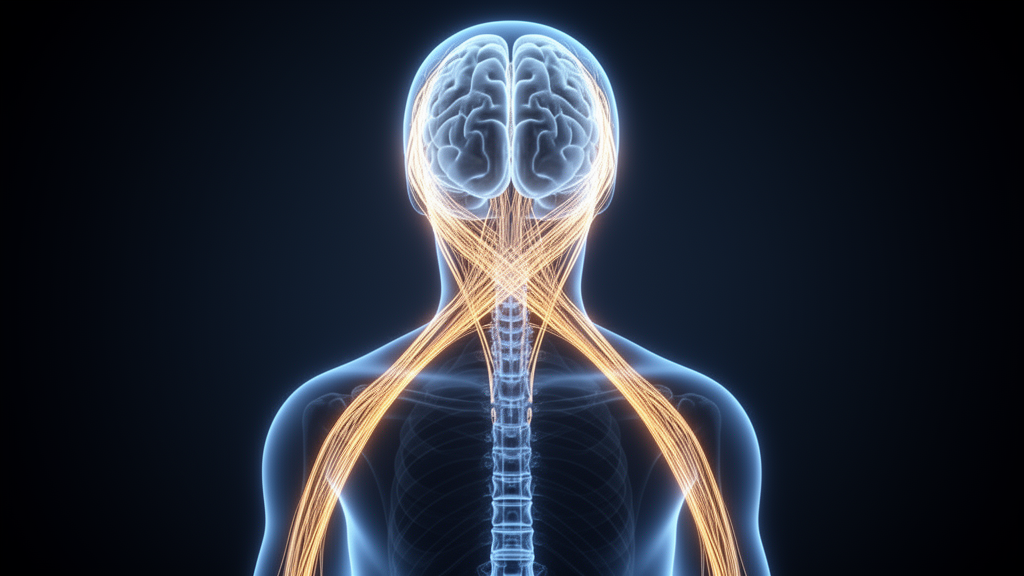
This crossed arrangement, known as contralateral organization, is not a quirk of human anatomy but a deep pattern shared across backboned animals. Now, in a guest editorial published in the journal Frontiers in Neuroanatomy, neuroscientists Daniel Wang and Martin Wessendorf of the University of Minnesota survey the major cross-wired circuits of four-limbed animals and advance a novel hypothesis about how this architecture came to be. Wessendorf is an associate professor emeritus whose research focuses on how the brainstem controls the function of neurons in the spinal cord, while Wang studies cognitive neuroscience and neurodegenerative diseases. Their proposal is striking in its simplicity: contralateral organization may not have evolved as an adaptation at all, but as a consequence of evolution building new circuits on top of an ancient visual blueprint.
The story the authors tell begins with the eyes. Because the lens of the eye inverts the image projected onto the retina, whatever lies in the right visual field strikes the left half of the retina, and whatever lies to the left strikes the right half. At the optic chiasm, the junction where the optic nerves meet, nerve fibers are sorted so that information from the right visual field is routed to the left half of the brain, with the mirror-image arrangement handling the left visual field. This sorting is not arbitrary; it is the first step in a chain of crossed connections that extends deep into the brain and, ultimately, down into the spinal cord. The primary early destination of these visual signals is an ancient midbrain structure called the optic tectum, known in mammals as the superior colliculus. The tectum generates rapid, automatic responses to what an animal sees, such as turning toward a sudden movement in the periphery.
Crucially, information from each visual field is mapped onto the opposite side of the tectum. The structure does not handle vision alone: in its deeper layers, somatosensory information, meaning physical sensations from the body, is mapped as well, in registration with the visual inputs. The optic tectum is found in every living vertebrate species, which makes it one of the oldest recognizable neuroanatomical structures in the vertebrate lineage. From sensation to the brain, the pattern of crossing continues along two major ascending routes that carry physical sensations from most of the body. The first is the dorsal column pathway, which mediates light touch, vibration, and the sense of body position. As its name suggests, it transmits sensations from the limbs and body by ascending the dorsal column in the spinal cord, where it connects to another neuron and crosses the midline in the brainstem, the region located just above the spinal cord, before continuing upward.
Notably, this pathway is absent in fishes but has been observed in amphibians, birds, mammals, and reptiles, demonstrating a highly conserved presence across tetrapods, the four-limbed vertebrates that include mammals, birds, reptiles, and amphibians. The second ascending route is the spinothalamic tract, which carries information about pain, itch, and some forms of touch from the body to the brain. It differs from the dorsal column pathway both in the sensations it conveys and in where it crosses: its fibers decussate within the spinal cord itself before ascending and conveying their sensory information to the thalamus, the part of the brain that sits just above the midbrain and processes much movement and sensory information. A contralaterally organized spinothalamic tract has been identified in birds, mammals, and reptiles, and even in some cartilaginous fishes such as sharks and rays. Together, these two pathways show that sensory traffic from the body reliably reaches the opposite side of the brain, a pattern repeated across a wide range of vertebrate lineages.
The authors then turn to the descending side of the conversation, examining three cross-wired pathways that control skeletal muscle. The first is the tectospinal pathway, the motor arm of the optic tectum. Because the tectum integrates visual, auditory, and somatosensory maps in register, it can trigger orienting movements toward salient events in any of these senses. The authors offer a familiar example: if you are about to enter a crosswalk and see an oncoming car from the corner of your eye, you turn towards it. This is one instance of how the tectospinal pathway mediates responses to visual stimuli, and it can mediate similar responses to auditory and tactile stimuli. Its motor component originates in the deepest layers of the optic tectum and projects its axons to the contralateral spinal cord. This pathway is present in most tetrapods and in some cartilaginous fishes.
The second descending route is the rubrospinal pathway, which is thought to facilitate limb control in animals. It is large in most mammals, originates in the red nucleus of the midbrain, and projects to the contralateral spinal cord. Like the tectospinal pathway, it has been reported to exist in tetrapods and some cartilaginous fishes. The third and most famous example is the corticospinal pathway, which in humans allows for fine motor control of the fingers and hands. This pathway is a special case, and its history reveals something unexpected about how our own dexterity may have arisen. All vertebrates possess a telencephalon, the front part of the brain that makes up most of its mass in humans, but only in mammals does the telencephalon contain a six-layered cerebral cortex, the tissue responsible for higher-order functions such as the integration of different sensations with memory, motor control, and language.
In most mammals, the corticospinal pathway travels from the cerebral cortex, projects to the part of the spinal cord where sensations enter, and modulates somatosensory input rather than driving movement. In primates, including humans, and in a few other dexterous species, the tract instead projects to the part of the spinal cord that controls the muscles, enabling, for instance, fine control of the fingers. Thus, depending on the species, the corticospinal pathway can be either a motor pathway or a pathway that modulates somatosensation. In both variants, the corticospinal tract crosses the brain midline. Wang and Wessendorf draw a provocative conclusion from this: humans appear to have obtained their fine motor control by co-opting a circuit whose ancestral function was somatosensory control, retaining the ancestral crossing while repurposing the output.
Putting the pieces together, the authors propose that evolutionarily ancient visual pathways biased the vertebrate central nervous system towards being organized contralaterally, at least in tetrapods. Because every living vertebrate has both an optic chiasm and a contralaterally organized optic tectum, they reason that Cambrian-era jawless fishes, which lived roughly 538 to 486 million years ago, likely possessed both structures as well. That inference is testable: the authors suggest it may be possible to examine fossilized remains of the skulls of those early fishes for evidence bearing on the hypothesis. If confirmed, the crossed arrangement of our sensory and motor systems would trace its origin to the visual organization of some of the earliest animals with backbones.
The deeper lesson the editorial draws concerns the character of evolution itself. Evolution is conservative in its innovations: it often retains and builds upon existing structures, pathways, and mechanisms rather than inventing new ones from scratch. On this view, the organization of the visual system, and in particular the crossed mapping of the optic tectum, may have defined the organizational principles required of the sensory and motor pathways that evolved later. Contralateral organization, in other words, need not have been selected for as an advantage in its own right; it may simply be what happens when a lineage of nervous systems is built on a visual foundation laid hundreds of millions of years ago. Evolution, as the authors put it, is an engineer that builds with the foundation and materials it has at hand.
The editorial, published on 27 October 2026, offers no new experiments of its own but reframes a familiar fact of neuroanatomy as an evolutionary inheritance. By tracing the crossed connections of vision, touch, pain, and movement through fishes, amphibians, reptiles, birds, and mammals, Wang and Wessendorf assemble a coherent account of why the right side of the brain speaks for the left side of the body. The answer they propose is neither elegant adaptation nor accident, but inheritance: a pattern set by the oldest visual circuitry in the vertebrate brain, followed faithfully by every sensory and motor pathway that came after it, and whose ultimate confirmation may lie buried in the skulls of Cambrian fishes.
Subject of Research: Evolutionary origin of contralateral (crossed) brain-body wiring in vertebrates
Article Title: Why does the right side of the brain control the left side of the body? An evolutionary history
Article References: Why does the right side of the brain control the left side of the body? An evolutionary history. Original publication (Bibliographic details unavailable.)
Image Credits: AI Generated
DOI: Not provided
Keywords: neuroanatomy, contralateral organization, optic tectum, optic chiasm, dorsal column pathway, spinothalamic tract, corticospinal tract, tectospinal pathway, rubrospinal pathway, tetrapods, evolution, Cambrian jawless fishes