In an observational investigation of patients diagnosed with chronic central serous chorioretinopathy, researchers at the Zhongshan Ophthalmic Center have identified specific choroidal vascular patterns and late-phase angiographic signs that independently correlate with the presence of secondary macular neovascularization. Central serous chorioretinopathy represents one of the most prevalent vision-impairing retinopathies following age-related macular degeneration, diabetic retinopathy, and retinal vein occlusion, primarily manifesting as local neurosensory retinal detachment in the macula. While acute presentations are frequently self-limiting with subretinal fluid reabsorbing within three to six months without causing major retinal structural compromise, the chronic form involves progressive alterations within the retinal pigment epithelium and underlying choroid. This persistent state carries a heightened risk of secondary macular neovascularization, a sight-threatening complication that typically impairs visual prognosis and prompts a definitive transition in clinical management from observation or laser photocoagulation to intravitreal anti-vascular endothelial growth factor injections.
To uncover vascular imaging biomarkers capable of refining risk stratification and guiding surveillance, the study team analyzed widefield indocyanine green angiography along with multimodal imaging protocols across 293 eyes from 212 patients evaluated between September 2022 and June 2025. The overall cohort comprised 164 male individuals, accounting for 77.36 percent of participants, reflecting the documented demographic distribution of the disease, with an average patient age of 49.24 years and a mean symptom duration of 32.76 months. Among the enrolled cohort, 96 eyes presented with secondary macular neovascularization, representing 32.76 percent of the sample, whereas 197 eyes showed no neovascular development. Eyes complicated by neovascularization were observed in significantly older individuals and demonstrated significantly worse visual acuity compared to uncomplicated eyes, confirming the adverse functional impact associated with choroidal neovascular remodeling in chronic disease courses.
Diagnostic assessment relied on standardized multimodal imaging, including color fundus photography, optical coherence tomography, widefield optical coherence tomography angiography, fundus fluorescein angiography, and widefield indocyanine green angiography. Within this protocol, macular neovascularization was diagnosed as present when eyes exhibited heterogeneous, flat, irregular pigment epithelium detachments on cross-sectional scans alongside neovascular flow signals detected on en face angiographic slabs extending from the outer retina to the choriocapillaris. In addition, ill-defined hyperfluorescent lesions displaying late leakage and staining on fluorescein and indocyanine green angiograms served as supportive diagnostic evidence. Among the 96 eyes affected by neovascularization, nearly one-third had previously undergone anti-vascular endothelial growth factor therapy, with a median intervention course lasting eight months and a median delivery of three therapeutic injections.
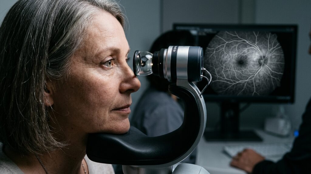
Widefield indocyanine green angiography permitted single-frame visualization of the choroidal vasculature across an expansive 102-degree field of view during early and mid-phases, supplemented by conventional 55-degree recordings extending past thirty minutes post-injection for late-phase analysis. Using a standardized nine-point peripheral fixation protocol encompassing the fovea, superior, superotemporal, temporal, inferotemporal, inferior, inferonasal, nasal, and superonasal positions, the investigators systematically documented the anatomical configuration of the vortex vein system. Physiological watershed zones partition normal choroidal drainage into four distinct quadrants, with the horizontal watershed traversing the fovea and optic disc, while the vertical watershed passes along the temporal margin of the papilla. The researchers quantified vortex vein ampullae per quadrant and tracked the emergence of dominant vortex veins whose terminal courses traversed the central fovea or optic disc.
Detailed comparative evaluation revealed distinct vascular architectures between the two clinical groups. While the total number of vortex vein ampullae and their quadrant-specific distributions did not differ significantly between eyes with and without neovascularization, asymmetric drainage patterns were detected significantly more frequently in eyes presenting with macular neovascularization, reaching 86.46 percent compared to 75.63 percent in uncomplicated cases. Furthermore, eyes with neovascularization demonstrated a higher frequency of solitary dominant vortex veins, occurring in 67.47 percent of cases versus 53.69 percent among eyes without neovascularization. When solitary dominant drainage vessels were identified, a superotemporal quadrant location emerged with significantly higher prevalence in the neovascular cohort compared to the non-neovascular group.
Venous remodeling was also evaluated through the identification of intervortex venous anastomoses, defined as two or more connecting vascular channels bridging adjacent quadrant drainage networks across horizontal or vertical physiological watersheds. The researchers categorized these collateral channels into macular, peripapillary, and peripheral subtypes based on their exact anatomical positions. Notably, while peripheral and peripapillary anastomoses showed comparable frequencies between groups, anastomotic vessels traversing the macular region were significantly enriched in eyes with neovascularization, observed in 60.42 percent of affected eyes compared to 46.70 percent in non-neovascular eyes. This localized prominence reinforces hypotheses that venous overload and hemodynamic congestion across the central macula trigger distinct vascular compensation mechanisms.
Late-phase angiographic evaluations focused on the identification and grading of age-related scattered hypofluorescent spots on late-phase indocyanine green angiography, an imaging feature that manifests as discrete dark spots during prolonged dye transit. These hypofluorescent alterations were documented far more frequently in eyes with macular neovascularization, appearing in 17.71 percent of neovascular cases compared to only 5.08 percent of non-neovascular eyes. While the qualitative presence of these spots differed markedly between cohorts, the severity grading—stratified according to macular, peripapillary, or diffuse posterior pole distribution—showed no statistically significant distinction, indicating that the mere appearance of this angiographic phenomenon carries primary clinical relevance.
To rigorously evaluate the relative contributions of these vascular features while accounting for bilateral eye correlation from individual patients, the investigators applied a generalized linear-mixed model. In multivariate analysis adjusted for patient age, sex, and duration of symptoms, three choroidal characteristics remained independently associated with the presence of secondary macular neovascularization: the presence of a superotemporal dominant vortex vein, macular intervortex venous anastomoses, and the detection of age-related scattered hypofluorescent spots on late-phase angiography. The authors noted that the statistical odds ratio for late hypofluorescent spots was accompanied by a wide confidence interval, an analytical result attributable to the relatively low overall frequency of this biomarker within the study population that warrants measured interpretation.
These statistical associations align with current mechanistic frameworks centered on the venous overload hypothesis in pachychoroid disorders. Because the temporal choroidal network drains high-volume blood flow from the macula against gravity along the superior pathway, venous engorgement in a dominant superotemporal vessel may generate sustained hemodynamic resistance, choriocapillaris compression, and localized capillary ischemia. Similarly, macular venous anastomoses reflect compensatory collateral formation in response to persistent choroidal congestion, although severe mechanical engorgement may further compound focal capillary hypoperfusion. Concurrently, late hypofluorescent spots may indicate retinal pigment epithelium dysfunction and the accumulation of lipoproteinaceous basal linear deposits, which impair oxygen diffusion across Bruch’s membrane and potentially stimulate local vascular endothelial growth factor release within a chronically hypoxic environment.
The study authors highlighted several methodological limitations inherent to their research design. Because the investigation was cross-sectional and retrospective, the temporal sequence between anastomotic channel remodeling and neovascular formation cannot be definitively proven, leaving open the possibility that macular anastomoses partially represent secondary adaptive responses to altered perfusion demands. Furthermore, proposed pathophysiological models linking angiographic venous overload to choriocapillaris ischemia remain theoretical frameworks requiring direct confirmation through histopathological investigations, and certain features like posterior vortex veins and late hypofluorescent spots exhibited low sample prevalence. Nevertheless, by demonstrating high interobserver agreement across widefield metrics, the findings establish tangible choroidal vascular biomarkers that support enhanced monitoring strategies for high-risk patients with chronic chorioretinopathy.
Subject of Research: Choroidal vascular biomarkers for macular neovascularization in chronic central serous chorioretinopathy
Article Title: Choroidal vascular biomarkers for macular neovascularization in chronic central serous chorioretinopathy: insights from widefield indocyanine green angiography
Article References: Hao, X., Pu, J., Gan, Y., Su, Y., Zhuang, X., He, G., Zhang, Y., Chen, X., He, Y., Zhang, G., Li, M., & Wen, F. (2026). Choroidal vascular biomarkers for macular neovascularization in chronic central serous chorioretinopathy: insights from widefield indocyanine green angiography. Eye and Vision, 13(1), Article 23. https://doi.org/10.1186/s40662-026-00494-0
Image Credits: AI Generated
DOI: 10.1186/s40662-026-00494-0
Keywords: central serous chorioretinopathy, macular neovascularization, indocyanine green angiography, choroidal vasculature, retina, ophthalmology, vortex vein, optical coherence tomography, Choroidal, vascular, biomarkers, macular