A preliminary preclinical pilot study evaluating an off-pump ascending aorta to left anterior descending artery bypass using internal mammary vein conduits in three Bama minipigs resulted in severe graft occlusion within four weeks. In the investigation conducted by researchers from Shanghai East Hospital and partner institutions, all three animal subjects demonstrated successful surgical graft placement with documented immediate intraoperative blood flow. However, subsequent coronary computed tomography angiography and histological evaluation at four weeks revealed complete absence of luminal contrast opacification alongside extensive occlusive structural remodeling. Mean luminal cross-sectional stenosis reached 97.4 percent across the most remodeled segments of the harvested grafts. The researchers developed this large-animal model to reproduce the anatomical configuration of clinical coronary artery bypass grafting, establishing that the off-pump procedure is technically feasible in swine while exhibiting a rapid occlusive phenotype. Nonetheless, the authors emphasized that this preliminary pilot investigation was not designed to establish model reproducibility, determine the exact temporal sequence of vessel closure, or isolate the precise biological mechanisms driving graft failure.
Coronary artery bypass grafting remains one of the most effective revascularization strategies for patients with severe coronary artery disease. Although arterial grafts provide superior long-term patency, the saphenous vein graft continues to be widely used because of its availability, length, and ease of harvest. However, vein graft failure remains a major limitation of coronary bypass surgery. Published clinical literature indicates that vein graft failure occurs in approximately 10 to 25 percent of grafts within the first year after surgery, and long-term patency decreases substantially over time, with only about half remaining patent at ten years. The pathological progression of graft failure encompasses early thrombosis, progressive intimal hyperplasia, adverse vascular remodeling, and accelerated atherosclerosis. Clinically, failed vein conduits are associated with recurrent myocardial ischemia, repeat revascularization, myocardial infarction, and adverse long-term cardiovascular outcomes. Therefore, elucidating the cellular mechanisms of graft failure and establishing reliable preclinical evaluation systems remain essential objectives in cardiovascular research.
Animal models are essential for studying the pathogenesis of vein graft failure and evaluating therapeutic interventions before clinical translation. Compared with small rodent models, large-animal models offer multiple translational advantages, particularly swine. The porcine cardiovascular system closely resembles human physiology in vascular dimensions, coronary anatomy, vessel wall structure, and hemodynamic characteristics. Furthermore, the comparatively large caliber of porcine vessels permits detailed histological, cellular, and molecular analyses from the same harvested graft specimen. Existing porcine vein graft investigations have predominantly relied on extra-coronary interposition models, such as grafting saphenous or jugular vein segments into the carotid circulation. Although carotid interposition exposes venous tissue to arterial pressure, it differs from an aortocoronary bypass in anatomical location, anastomotic geometry, target vessel, and downstream vascular bed. An ascending aorta to coronary bypass allows researchers to observe venous behavior directly within the coronary circulation and evaluate hemodynamic interactions with native coronary flow.
In swine, the great saphenous vein can be small and closely associated with the saphenous artery, complicating harvest for coronary grafting. Addressing this limitation, the authors evaluated a free internal mammary vein in an off-pump ascending aorta to left anterior descending artery configuration. Harvested through the same median sternotomy required for cardiac exposure, the internal mammary vein provides practical length and caliber. However, it is not a direct structural equivalent of the human saphenous vein; its thin wall and thoracic environment may confer different mechanical and biological responses under arterial pressure. Approved under protocol 20250303 at the Laboratory Animal Center of Shanghai Jiao Tong University Agricultural and Biological Experimental Practice Farm, the study enrolled three female Bama minipigs aged six months, weighing 30.3 ± 1.5 kilograms. This small cohort was selected pragmatically in accordance with the reduction principle to assess surgical feasibility and endpoint tissue characteristics while limiting large-animal use.
Anesthesia was induced with intramuscular tiletamine hydrochloride and zolazepam hydrochloride at 7 milligrams per kilogram, followed by endotracheal intubation and mechanical ventilation with 2.0 to 2.5 percent inhaled isoflurane. Following median sternotomy, the left internal mammary vein was dissected from its distal bifurcation proximally toward the thoracic inlet, preserving the accompanying artery. Across the three animals, harvested veins yielded a mean length of 10.8 ± 0.7 centimeters and a mean caliber of 4.1 ± 0.2 millimeters. Before implantation, a one-centimeter distal segment was excised and preserved as a normal within-animal tissue comparator. Side branches were clipped with titanium clips, and conduits were preserved in lactated Ringer’s solution without deliberate distension.
Systemic anticoagulation was initiated using intravenous heparin at 1 milligram per kilogram to achieve an activated clotting time exceeding 300 seconds. Both vascular connections were constructed end-to-side on a beating heart without cardiopulmonary bypass. The native distal end of the internal mammary vein was anastomosed to the ascending aorta with continuous 6-0 polypropylene sutures, while the native proximal end was connected to the left anterior descending artery using continuous 7-0 polypropylene sutures. Because the harvested vein measured approximately 4 millimeters in caliber while the recipient coronary artery measured approximately 2 millimeters, the coronary arteriotomy length was extended to approximately three times the target vessel diameter. The native left anterior descending artery was left unobstructed without proximal narrowing or ligation.
Following completion of both anastomoses, heparin was neutralized with protamine sulfate, and graft function was immediately assessed using transit-time flow measurement. Intraoperative hemodynamic recordings confirmed graft patency in all three animals, with individual mean graft flows measuring 18, 25, and 21 milliliters per minute, resulting in a cohort mean of 21.3 ± 3.5 milliliters per minute. The corresponding pulsatility indices were 4.1, 3.6, and 3.8, averaging 3.8 ± 0.3, while diastolic fractions were 52, 56, and 53 percent, averaging 53.7 ± 2.1 percent. Total operative time averaged 134.3 ± 8.6 minutes, with zero perioperative mortality, no ventricular fibrillation, and no requirement for cardiopulmonary bypass conversion. Temporary chest drainage tubes were placed under negative pressure suction, the sternum was reapproximated with stainless-steel wires, and layered wound closure was completed. After restoring spontaneous breathing, all animals were extubated on the day of surgery and received oral aspirin daily at 100 milligrams per day until euthanasia at four weeks.
At four weeks postoperatively, all three minipigs underwent coronary computed tomography angiography to evaluate graft patency, with images independently assessed by two experienced cardiovascular surgeons. The imaging demonstrated complete lack of luminal contrast opacification throughout all three bypass grafts, classifying every graft as occluded. While three-dimensional volume-rendered computed tomography delineated the adjacent native coronary vasculature, occluded, non-opacified venous conduits could not be reliably visualized on surface reconstructions alone. The authors emphasized that these imaging findings established the loss of detectable lumen patency by four weeks, but could not identify the precise time point when occlusion began. In particular, intraoperative transit-time flow measurement confirmed only immediate postoperative patency and could not rule out early thrombosis occurring shortly after surgery. Furthermore, radiological non-opacification on angiography describes macroscopic contrast passage and must be distinguished from microscopic cross-sectional stenosis, as microscopic residual channels may persist without permitting visible angiographic opacification.
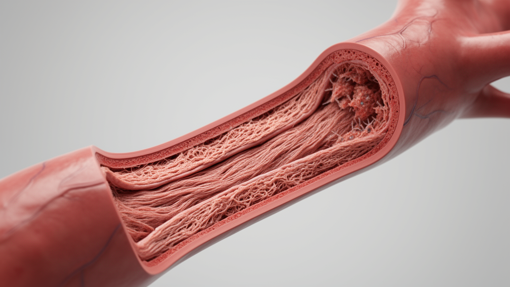
Following angiography, animals were re-anesthetized and humanely euthanized using intravenous potassium chloride at 100 milligrams per kilogram, and the bypass grafts were harvested en bloc. Macroscopic resternotomy inspection revealed marked vessel thickening and loss of the thin-walled venous morphology seen during implantation. Histological cross-sections stained with hematoxylin and eosin demonstrated extensive fibrocellular tissue accumulation causing near-total luminal obliteration. Morphometric analysis of transverse sections selected from regions displaying the greatest gross remodeling showed cross-sectional stenosis rates of 97.0 percent in pig 1, 99.3 percent in pig 2, and 95.8 percent in pig 3, yielding a cohort mean stenosis of 97.4 ± 1.8 percent. The measured intimal area expanded markedly from 662,764.4 ± 83,923.6 square micrometers in native veins to 4,883,748.6 ± 1,035,710.7 square micrometers in four-week explanted grafts. The investigators stressed that these measurements were deliberately taken from the most severely remodeled regions to characterize extreme lesion progression and do not represent whole-graft averages.
Extracellular matrix and cellular remodeling were further evaluated using specialized histology and confocal immunofluorescence. Masson’s trichrome staining demonstrated substantial collagen accumulation within remodeled grafts compared with native controls, while Elastic Van Gieson staining revealed severe disruption and fragmentation of elastic fiber architecture. Dual immunofluorescence showed dense accumulation of alpha-smooth muscle actin-positive cells throughout the occlusive lesions, contrasted with the media-restricted distribution in native veins. In addition, the lesions contained numerous irregular CD31-positive endothelial-lined channels, a pattern compatible with vascular reorganization or recanalization of organized thrombus. Discussing study limitations, the authors highlighted the small three-animal sample size, the absence of serial early imaging to determine the exact onset of occlusion, and the presence of competitive flow from the unobstructed native coronary artery. Consequently, the authors concluded that larger cohorts and earlier surveillance are required before this off-pump aortocoronary model can serve as an established platform for testing preventive biomaterials or therapeutic interventions.
Subject of Research: Preclinical assessment of off-pump aortocoronary vein graft failure in swine
Article Title: Development and preliminary characterization of a rapid porcine coronary vein graft failure model
Article References: Zhu, P., Wu, W., Cao, D., Huang, Q., Lu, H., Xue, B., Ling, Y., Liu, J., & Liu, J. (2026). Development and preliminary characterization of a rapid porcine coronary vein graft failure model. Frontiers in Bioengineering and Biotechnology, 14, Article 1986776. https://doi.org/10.3389/fbioe.2026.1986776
Image Credits: AI Generated
DOI: 10.3389/fbioe.2026.1986776
Keywords: coronary artery bypass grafting, vein graft failure, Bama minipigs, internal mammary vein, off-pump coronary bypass, left anterior descending artery, aortocoronary bypass, intimal hyperplasia, transit-time flow measurement, coronary computed tomography angiography, luminal stenosis, vascular remodeling, alpha-smooth muscle actin, CD31 endothelial marker, elastic fiber architecture, extracellular matrix remodeling, preclinical porcine models