A seemingly routine Arabidopsis mutant line has turned out to carry a hidden piece of extra DNA that changes how the plant grows when light is scarce. Researchers studying mutants of mitochondrial malate metabolism discovered that the unexpectedly severe phenotype of a double mutant was not caused by the intended gene knockouts alone, but by a large inverted duplication lurking next to one of the T-DNA insertion sites. The finding, published in Plant Cell Reports, is a cautionary tale for the thousands of laboratories that rely on T-DNA insertion lines to work out what genes do.
The team, led by María del Pilar Martinez and Veronica G. Maurino at the University of Cologne together with colleagues including Boas Pucker, set out to examine how plants cope when two routes of mitochondrial malate metabolism are knocked out simultaneously. They combined a mutation in MDH1, which encodes the predominant mitochondrial malate dehydrogenase of green tissues, with defects in ME1 and ME2, the two genes whose products form the heterodimeric NAD-dependent malic enzyme. Because NAD-ME acts mainly as a heterodimer in vivo, the researchers expected a double mutant lacking only ME2 to look essentially the same as a triple mutant lacking ME1 and ME2 both.
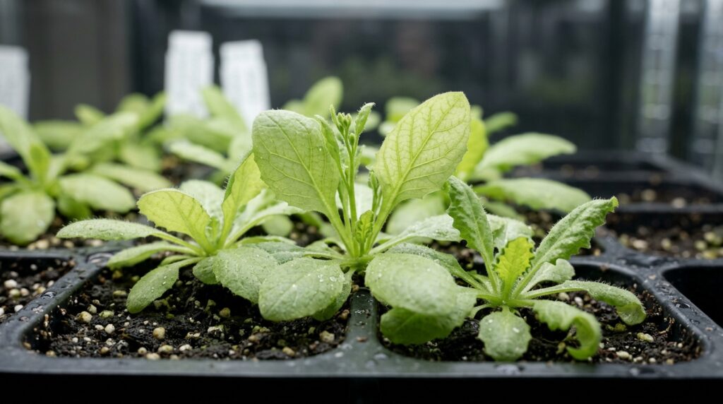
Under long days at normal light intensity of 120 micromol photons per square metre per second, that expectation held: no visible differences separated the mutants from wild type. The surprises appeared when the plants were pushed into energy-limiting conditions. Under short days at normal light, the double mutants already grew less than wild type, while the triple mutant remained largely indistinguishable from it. Under short days at low light of 60 micromol photons per square metre per second, the double mutants became dramatically smaller than wild type, with pale green leaves, and failed to flower altogether. Strikingly, the triple mutant, which carries one more knocked-out gene, stayed consistently larger and less chlorotic than either double-mutant line.
That inversion of expectation, more missing genes producing a milder plant, made no sense if the phenotype simply reflected the loss of MDH1 and NAD-ME function. Measurements of photosynthetic performance deepened the puzzle. In the double mutant grown under short days at low light, the maximum quantum efficiency of photosystem II fell to roughly half the value seen under normal light, the effective quantum yield and electron transport rate dropped close to zero, and non-photochemical quenching rose to about twice its normal-light level. Transmission electron microscopy revealed chloroplasts with markedly thicker, hyperstacked grana thylakoids and an increased number of plastoglobules, features associated with remodeling of the photosynthetic apparatus and with stress.
The first clue that something beyond the intended mutations was at play came from biochemistry. In wild-type mitochondrial extracts, NAD-ME activity was readily detectable, but activity was below the detection limit in both the double and triple mutants. Immunoblotting found a clear ME1 band in wild type but no immunoreactive signal in either mutant line, and mass-spectrometry-based proteomics failed to detect any ME1-derived peptides in the mutants. In other words, both genotypes effectively lacked heterodimeric NAD-ME, so the harsher phenotype of the double mutant could not be explained by residual ME1 activity.
To look for hidden genetic lesions, the researchers turned to whole-genome sequencing with Oxford Nanopore long reads. Local assemblies confirmed the T-DNA insertions at the MDH1 locus on chromosome 1 and the ME2 locus on chromosome 4. But the structural-variant analysis also uncovered something unplanned: in both independently generated double-mutant lines, an approximately 137-kilobase segment immediately downstream of the MDH1 T-DNA insertion site had been duplicated and inserted back in an inverted orientation. The duplicated interval, spanning positions from roughly 19.86 to 19.99 million base pairs on chromosome 1 of the TAIR10 reference, contains 38 annotated genes. Crucially, the same duplication was absent from the triple mutant.
Genetic evidence tied the severe phenotype to this structural variant rather than to the gene knockouts. When the researchers re-examined the original mdh1.1 single-mutant population under short days at low light, it segregated into distinct phenotypic classes, including wild-type-like plants and small, yellowish individuals, exactly what one would expect if some members of the population carried the duplication and others did not. An independent MDH1 insertion allele, mdh1.2, showed a uniform wild-type-like phenotype, and double mutants built on mdh1.2 did not reproduce the severe growth defect. The researchers reconstructed how this happened: the original mdh1.1 parental population likely contained MDH1 alleles that differed in whether the adjacent duplication was present, and because all mutant selection was done under long days at normal light, where no visible phenotype appears, the duplication-carrying allele was picked by chance when the double mutants were made, while a duplication-free allele was picked by chance for the triple mutant.
The duplication was anything but silent. RNA sequencing showed that 32 of the 38 genes in the interval produced detectable transcripts, and their abundance rose most strongly around the dark-to-light transition under short days at low light, when 19 of the 32 genes were significantly upregulated relative to wild type. Proteomics detected only five of the 38 encoded proteins: phosphoenolpyruvate carboxylase 1, or PEPC1; the mitochondrial sulfur dioxygenase ETHE1; the oxidative-stress protein DJ1B; the UDP-L-rhamnose biosynthetic enzyme RHM2; and fatty-acid-binding protein 3. Of these, PEPC1 stood out, accumulating consistently in the duplication-carrying double mutant while staying near wild-type levels in the triple mutant. PEPC enzymatic activity was significantly higher in the double mutant under low light, and immunoblotting confirmed a stronger PEPC1 band of the expected 110 kilodaltons. The PEPC1 promoter also carries light-responsive elements, including a G-box and a GT1-motif, offering a plausible regulatory basis for the condition-dependent expression.
The metabolic consequences fit a coherent picture. Under low light the double mutants showed a reduced carbon-to-nitrogen ratio, driven by lower total carbon and higher total nitrogen, along with significantly elevated leaf ammonium, changes that were absent under normal light. Untargeted gas-chromatography mass-spectrometry profiling revealed a coordinated accumulation of oxaloacetate-derived amino acids: aspartate, lysine, threonine and isoleucine rose roughly fourfold, while asparagine increased fifteen- to twentyfold relative to wild type. None of this was seen in the triple mutant. Because PEPC1 produces oxaloacetate for anaplerotic carbon fixation, the authors propose that increased PEPC1 abundance may channel carbon toward nitrogen-rich storage compounds such as asparagine when energy is limiting, perturbing carbon and nitrogen balance. They are careful, however, to call PEPC1 a candidate contributor rather than the sole cause: the duplicated interval contains many other genes involved in signaling, proteostasis and stress responses, and an interaction between the duplication and the loss of MDH1 cannot be excluded.
The broader lesson reaches well beyond one metabolic pathway. T-DNA insertion mutagenesis underpins enormous indexed collections such as SALK, GABI-Kat, SAIL and WISC, and insertion events are frequently complex, involving truncated borders, tandem repeats, multiple copies and plasmid backbone sequences. Large deletions, inversions, duplications and translocations linked to integration events have been reported repeatedly, and recent studies have traced puzzling mutant phenotypes to megabase-scale inversions and reciprocal translocations rather than to the annotated target gene. The authors argue that when a mutant phenotype deviates from genetic expectation, particularly in an environment-dependent way, structural validation of insertion loci by long-read sequencing should be a standard prerequisite before drawing conclusions about gene function, especially as Agrobacterium-mediated DNA transfer remains a workhorse of plant genome editing.
Subject of Research: A hidden T-DNA-linked inverted duplication that alters gene dosage and exacerbates light-dependent phenotypes in Arabidopsis mutants
Article Title: A hidden T-DNA-linked inversion-duplication is associated with a light-dependent Arabidopsis mutant phenotype
Article References: del Pilar Martinez, M., de Oliveira, J. A. V. S., Nica, I., Ditz, N., Zheng, K., Wewer, V., Metzger, S., Westhoff, P., Eubel, H., Finkemeier, I., Schwarzländer, M., Pucker, B., & Maurino, V. G. (2026). A hidden T-DNA-linked inversion-duplication is associated with a light-dependent Arabidopsis mutant phenotype. Plant Cell Reports, 45(11), Article 350. https://doi.org/10.1007/s00299-026-04030-1
Image Credits: AI Generated
DOI: 10.1007/s00299-026-04030-1
Keywords: Arabidopsis thaliana, T-DNA insertion, structural variation, inverted duplication, MDH1, NAD-malic enzyme, PEPC1, gene dosage, carbon-nitrogen metabolism, long-read sequencing, mutant phenotyping, functional genomics