Surgeons have demonstrated the feasibility and safety of utilizing the ligamentum teres hepatis as a pedicled, autologous buttress for crural reinforcement during laparoscopic paraesophageal hernia repair in a patient with a prior sleeve gastrectomy. Documented as a case report and surgical video article published in Innovative Surgical Trends by Salma Khan, Naveed Haroon, Nolberto Jaramillo, Racheal Barnes, and Ashutosh Kaul, the report details an operative approach designed to address complex hiatal disruption without relying on prosthetic mesh. The authors achieved successful reduction of the herniated stomach, restored intra-abdominal esophageal length, and reinforced the posterior cruroplasty, resulting in an uneventful recovery with resolved reflux symptoms and no early recurrence.
Sleeve gastrectomy stands as the most commonly performed bariatric procedure worldwide, yet postoperative gastroesophageal reflux disease and secondary paraesophageal hernias can arise or progressively deteriorate following the initial resection. Addressing a paraesophageal hernia in this surgical population presents substantial technical difficulty because prior anatomical alteration precludes standard reconstructive techniques. The absence of the gastric fundus eliminates the possibility of performing a traditional fundoplication, while placing prosthetic mesh at the hiatus remains highly controversial due to recognized risks of erosion, stricture formation, and localized infection. Although conversion to a Roux-en-Y gastric bypass represents an alternative consideration in selected patients, the investigators note that such revisional bypass carries additional operative risks.
The reported procedure involved a 47-year-old female patient with a history of morbid obesity, characterized by a preoperative body mass index of 44 kg/m², who had undergone a laparoscopic sleeve gastrectomy three years earlier with no paraesophageal hernia noted at that time. Her past medical history also included chronic lower back pain and spondylolysis. Over the intervening years, the patient experienced progressive gastroesophageal reflux symptoms, persistent epigastric discomfort, and postprandial intolerance that continued to worsen despite receiving maximal medical therapy. Preoperative diagnostic workup, consisting of upper endoscopy and cross-sectional imaging, revealed a 10 cm paraesophageal hernia with substantial herniation of the gastric sleeve into the mediastinal cavity, prompting the recommendation for revisional surgical repair after obtaining informed consent.
The operation was conducted laparoscopically under general anesthesia with the patient positioned in a supine, reverse Trendelenburg orientation and trocars placed in a triangulated configuration across the upper abdomen. Upon abdominal entry, the surgical team encountered dense adhesions resulting from the prior bariatric intervention, which were meticulously lysed to achieve clear visualization of the diaphragmatic hiatus. The surgeons circumferentially dissected the hernia sac and completely reduced its contents from the mediastinum back into the peritoneal cavity. Throughout the mediastinal dissection, both the anterior and posterior vagus nerves were identified and carefully preserved, while dissection along the distal esophagus continued until adequate intra-abdominal esophageal length was established without structural tension.
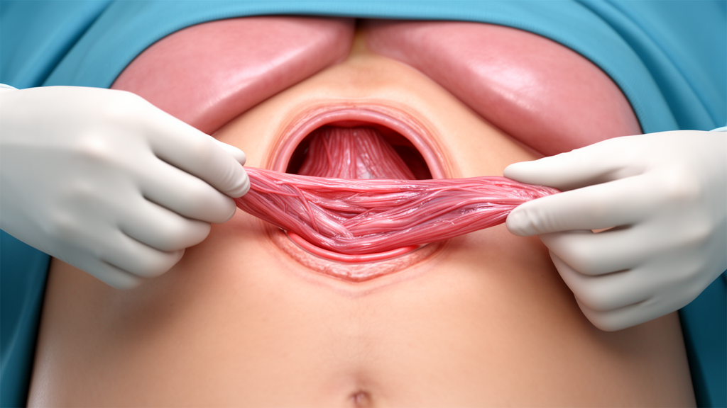
Following full mediastinal reduction and esophageal mobilization, the surgeons performed a posterior cruroplasty utilizing interrupted, nonabsorbable sutures to close the enlarged diaphragmatic defect. To reinforce the crural closure without implanting synthetic material, the surgical team mobilized the ligamentum teres hepatis from the anterior abdominal wall while deliberately preserving its native vascular pedicle. The mobilized, vascularized ligament was then rotated posteriorly into the hiatus and anchored over the primary crural suture line using interrupted sutures. This maneuver created a living autologous buttress directly over the repaired crus, with no fundoplication constructed due to the altered sleeve anatomy, and the procedure concluded with achieved hemostasis and zero intraoperative complications.
The patient’s postoperative course proceeded without adverse events. On postoperative day one, an objective contrast swallow examination was administered, demonstrating normal, unobstructed passage of oral contrast into the stomach with no radiographic evidence of contrast extravasation, leak, or recurrent herniation. Following this favorable imaging study, the patient’s intake was advanced to a liquid diet, which was tolerated well without episodes of nausea or vomiting. The patient was subsequently discharged home in stable clinical condition. During early outpatient follow-up evaluations, the patient documented marked symptomatic relief, reporting complete resolution of her preoperative complaints, absence of reflux symptoms, and no clinical indications of early hernia recurrence.
In evaluating the broader implications of the case, the authors emphasize that utilizing the ligamentum teres hepatis provides an autologous, well-vascularized, readily accessible tissue buttress that avoids the serious foreign-body complications associated with permanent prosthetic mesh at the hiatus. While prior literature has validated the safety and feasibility of ligamentum teres reinforcement in complex hiatal repairs, this report confirms its specific applicability to foregut anatomy altered by prior sleeve gastrectomy. Nonetheless, the authors acknowledge that because this report reflects a single operative case with short-term clinical evaluation, extended long-term follow-up remains necessary to fully determine procedural durability, late structural integrity, and definitive hernia recurrence rates in revisional bariatric surgery.
Subject of Research: Laparoscopic paraesophageal hernia repair using an autologous ligamentum teres buttress after sleeve gastrectomy
Article Title: Laparoscopic paraesophageal hernia repair using ligamentum teres buttress after sleeve gastrectomy
Article References: Khan, S., Haroon, N., Jaramillo, N., Barnes, R., & Kaul, A. (2026). Laparoscopic paraesophageal hernia repair using ligamentum teres buttress after sleeve gastrectomy. Innovative Surgical Trends, 2(1), Article 19. https://doi.org/10.1007/s44414-026-00032-4
Image Credits: AI Generated
DOI: 10.1007/s44414-026-00032-4
Keywords: paraesophageal hernia, sleeve gastrectomy, ligamentum teres hepatis, crural reinforcement, posterior cruroplasty, laparoscopic repair, autologous buttress, mesh-free repair, gastroesophageal reflux disease, foregut surgery, hiatal hernia, bariatric surgery, mediastinal dissection, upper endoscopy, contrast swallow study, vagus nerve preservation, revisional surgery, case report