Neuroscientists at King’s College London have uncovered compelling new evidence highlighting the profound impact early visual environments have on the development of the eye and consequent behavioral responses in zebrafish. In the initial five days of life— a critical developmental window—the visual surroundings influence not only the architectural complexity of retinal neurons but also their electrical signaling patterns. These perceptual alterations extend beyond mere cellular morphology and directly affect behavioral preferences associated with visual stimuli, a discovery that challenges long-standing assumptions about the immutability of retinal structure early in life.
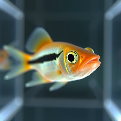
This groundbreaking research focused on zebrafish exposed to distinct visual settings characterized by either horizontal or vertical stripes. By carefully controlling these environmental variables during their formative phase, researchers observed striking differences in the dendritic morphology of retinal ganglion cells, the neurons responsible for transmitting visual information from the eye to the brain. The stripes were deliberately chosen due to their saliency as fundamental visual patterns that animals use to interpret complex scenes, such as faces, where vertical and horizontal lines form meaningful cues.
The study employed state-of-the-art microscopy to image neuronal structures at high resolution, revealing that fish nurtured in horizontally striped environments developed neurons with unique shapes and response properties compared to those exposed to vertical stripes. More importantly, electrophysiological recordings demonstrated functional plasticity—neuronal responses were biased to favor orientations that the fish had been exposed to during this early developmental period. This activity-dependent remodeling points to a previously unrecognized degree of adaptability within the retina itself, a sensory organ historically considered static and hardwired.
To bridge these physiological alterations with behavioral outcomes, the researchers devised an innovative virtual reality system in collaboration with the University of Konstanz. This setup allowed real-time tracking of zebrafish movement, testing their innate preference to swim toward stripes aligned parallel to their body axis. Remarkably, fish raised in horizontally striped surroundings exhibited a diminished preference for parallel stripes, indicating a disruption in their ability to discern orientation cues. Conversely, those raised amidst vertical lines preserved this instinctual behavior, reinforcing the link between retinal development and visual-guided actions.
The findings provoke a reevaluation of classical neuroscience models, which traditionally ascribed sensory plasticity chiefly to changes occurring within the cerebral cortex and other higher-order brain regions. Professor Robert Hindges, senior author and expert in developmental neurobiology, expresses surprise at discovering plasticity manifesting at such an early sensory stage. The retina, typically conceptualized as mere “hardware” responsible for initial light detection, is now emerging as a dynamic processor influenced by environmental exposure.
This paradigm shift implies that visual pre-processing begins not solely in the brain but originates within the retina, shaping perceptual frameworks even before cortical involvement. Consequently, environmental factors encountered during critical developmental windows can recalibrate fundamental sensory circuits, tailoring neural architectures to optimize processing for prevailing visual features. Such plasticity provides evolutionary advantages by enabling organisms to adapt to their habitats, fine-tuning sensory systems for efficient navigation and survival.
Further validating the retinal basis of these behavioral changes, genetic manipulation experiments selectively inhibited retinal plasticity. Under these conditions, zebrafish from both environmental groups exhibited indistinguishable behaviors, demonstrating conclusively that retina-specific alterations underpin the observed differences in stripe preference. This pinpointed the biological locus of plasticity, distinguishing it from potential brain-mediated influences.
These insights have broad implications extending beyond zebrafish, suggesting that the visual environments experienced during early development might influence perceptual systems across species, including humans. Similar mechanisms may account for variations in how individuals from diverse cultural or environmental backgrounds perceive optical illusions and spatial features. Understanding retinal plasticity opens prospects for novel therapeutic interventions targeting sensory processing disorders, by manipulating environmental exposure or harnessing intrinsic neuronal adaptability.
The research was facilitated by a multidisciplinary team integrating expertise in neuroscience, bioengineering, and behavioral science, equipped with cutting-edge virtual reality tracking and advanced imaging techniques. This synergy allowed a comprehensive exploration from molecular to behavioral scales, setting a new benchmark for studying sensory system development.
Funded predominantly by prestigious institutions such as the Medical Research Council, Leverhulme Trust, Biotechnology and Biological Sciences Research Council, and the Emmy Noether Program, this study was published in the esteemed journal Neuron on June 23, 2026. It offers a vital contribution to the growing recognition of sensory plasticity and its profound role in shaping perception and behavior.
Dr. Phoebe Reynolds, lead author, who conducted this research during her doctoral studies at King’s College London, now furthers her work as a postdoctoral researcher at the Friedrich Miescher Institute of Biomedical Research in Basel, Switzerland. This continuation signals vibrant ongoing explorations into the interplay between genetic programming and environmental experience in sensory system development.
In summary, these findings revolutionize our understanding of visual development by demonstrating that the retina is not a passive conduit but an active site of experience-dependent plasticity. Early visual environments sculpt the neural architecture and function of the retina, which in turn governs critical behaviors. This study shines a light on the fundamental processes orchestrating sensory perception, opening new avenues for research in developmental neuroscience, vision science, and the broader biological sciences.
Subject of Research: Animals
Article Title: Early visual experience elicits cellular and functional plasticity in the retina and alters behavior
News Publication Date: 23-Jun-2026
Web References:
https://doi.org/10.1016/j.neuron.2026.05.001
References:
Reynolds et al., Early visual experience elicits cellular and functional plasticity in the retina and alters behavior, Neuron, 2026.
Image Credits:
Francesca Greenstreet, King’s College London (videography); Reynolds et al. and Dr Paride Antinucci (microscopy).
Keywords:
Developmental neuroscience, Sensory perception, Perception, Visual perception, Systems biology, Neurophysiology, Vision, Neuroscience, Perceptual processes, Virtual reality
Tags: behavioral responses to visual cuescritical developmental window in zebrafishearly life visual stimuli effectshorizontal vs vertical stripe exposureneuronal electrical signaling patternsneuroscience of visual perceptionretinal ganglion cell morphologyretinal structure plasticityvisual environment impact on retinal neuronszebrafish eye developmentzebrafish virtual reality experimentzebrafish visual system research