
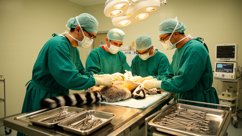
A recent clinical case study has identified the first documented instance of a vaginal fibroepithelial polyp in a non-human primate, providing significant new insights into reproductive tract health for wildlife veterinary medicine. The patient, a subadult female ring-tailed lemur (Lemur catta) housed at the Greens Zoological Rescue and Rehabilitation Centre in India, was initially presented with a recurrent, soft-tissue mass intermittently protruding from the vulva. However, the subsequent detailed examination revealed the condition was actually a localized, well-defined, pedunculated growth rather than the circumferential tissue protrusion typically associated with prolapse.
To ensure a precise diagnosis, the medical team performed a comprehensive reproductive tract evaluation while the animal was under general anesthesia. Utilizing a vaginal speculum and vaginoscopy, clinicians observed that the lesion originated from the left lateral vaginal wall, remaining strictly confined to that area. The examination confirmed that the cervix was closed, smooth, and anatomically normal, with no evidence of cervical or uterine involvement. This distinction proved critical, as it allowed the team to rule out more severe reproductive tract disorders and plan a targeted surgical intervention that minimized unnecessary trauma to the animal’s reproductive anatomy.
The surgical management involved the excision of the pedunculated mass using an advanced bipolar LigaSure™ vessel-sealing device. This technology allowed for the precise transection of the lesion’s stalk while maintaining excellent control of bleeding and reducing damage to the surrounding healthy vaginal tissue. The entire procedure, from induction to the animal’s smooth recovery, took approximately 50 minutes, with the surgical portion lasting about 30 minutes. Following the surgery, the animal received a multimodal pain management regimen including tolfenamic acid and tramadol, alongside oral amoxicillin–clavulanic acid to prevent secondary bacterial infections.
Histopathological analysis provided the definitive diagnosis of a benign vaginal fibroepithelial polyp. Microscopic evaluation revealed a mass composed of hyperplastic stratified squamous epithelium supported by a proliferative fibrovascular stromal core. The presence of focal acanthosis and finger-like epidermal projections into the underlying stroma, combined with the absence of cellular atypia, anisokaryosis, anisocytosis, or infiltrative growth, confirmed the benign nature of the lesion. While the subepithelial stroma exhibited widespread myxomatous expansion and scattered inflammatory cells, likely resulting from chronic mechanical irritation during the mass’s intermittent protrusion, there were no indications of neoplastic transformation.
The findings in this lemur share striking similarities with vulvovaginal fibroepithelial polyps frequently reported in human gynecological pathology. In human medicine, these lesions are often associated with hormonal influences—particularly estrogen and progesterone—and are frequently observed during pregnancy or in patients undergoing hormone therapy. Although the veterinary team was unable to conduct hormonal receptor analysis in this case to confirm a direct hormonal association, the morphology of the polyp and its clinical presentation align with established human clinical data, suggesting that similar benign stromal proliferative lesions may exist across various primate species.
Comparing these results to existing veterinary literature highlights the rarity of such conditions in non-human primates. While vaginal fibrous polyps have been documented in canine medicine, where they are also managed via surgical excision, reports within the order Primate remain extremely scarce. A prior case of a fibroepithelial polyp involving the ureter of an owl monkey exists in the literature, but this report represents the first known occurrence within the female reproductive tract of a prosimian, filling a notable gap in zoo and wildlife reproductive medicine records.
The success of the treatment was confirmed through long-term monitoring. After seven days of recovery, the lemur was safely reintroduced to her social group, where she resumed normal activity and behaviors. A routine annual screening conducted one year later, which included a follow-up vaginal endoscopy, showed that the surgical site had healed completely with no signs of recurrence. This positive outcome reinforces the efficacy of complete surgical excision for managing these benign lesions and highlights the importance of thorough diagnostic protocols when dealing with vulvar protrusions in wildlife species.
Clinical practitioners emphasize that this case serves as an important reminder not to assume all protruding vulvar masses in lemurs are vaginal prolapses. The case study demonstrates that detailed physical examinations and endoscopic evaluations are essential for achieving an accurate diagnosis and avoiding inappropriate treatment. By documenting this rare occurrence, researchers aim to improve the broader understanding of reproductive health in managed ring-tailed lemur populations, noting that while this report provides valuable comparative pathological information, further studies involving more cases will be necessary to fully elucidate the pathogenesis and potential hormonal associations of these polyps in primates.
The clinical background and captive environment provide vital context regarding reproductive monitoring in prosimians. Ring-tailed lemurs, classified scientifically as Lemur catta within the family Lemuridae, are diurnal primates native to Madagascar, averaging 2.2 kilograms in body weight. They inhabit dry deciduous forests, scrublands, and rocky landscapes across southern and southwestern Madagascar, living in social troops. The International Union for Conservation of Nature lists the species as Endangered due to habitat loss, deforestation, hunting, and the illegal pet trade. Ring-tailed lemurs reach sexual maturity between two and a half and three years of age, featuring a bicornuate uterus with two distinct horns. Their reproduction is strictly seasonal and regulated by photoperiod, meaning reproductive tract integrity is vital for copulation, pathogen exclusion, and parturition. The 2.3-kilogram subadult female in this case had no history of parturition. She lived in a stable group of six lemurs at Greens Zoological Rescue and Rehabilitation Centre in Jamnagar, Gujarat, India, alongside an adult male, an adult female, two juvenile males, and a subadult female. Their thirty-by-thirty-by-fifteen-foot enclosure offered indoor and outdoor paddocks with dense vegetation. The animals received fresh bamboo leaves, tamarind leaves, assorted vegetables, and Mazuri LowFe low-iron pellets to prevent hepatic iron storage disease. Positive reinforcement conditioning permitted stress-free transport to the hospital.
Preoperative clinical assessments established that the patient was systemically healthy before receiving anesthesia. Femoral vein blood samples analyzed for standard hematology and serum biochemistry yielded values within normal physiological intervals. Food was withheld for twelve hours and water for five hours prior to the procedure. Sedation was achieved with an intramuscular injection containing medetomidine at 0.03 milligrams per kilogram and ketamine at 3 milligrams per kilogram. Lateral recumbency occurred smoothly within six minutes, without alpha-2 agonist adverse effects such as vomiting, severe bradycardia, or hypotension. Anesthesia maintenance relied on isoflurane delivered between 0.5 and 4 percent in oxygen. Intravenous catheterization maintained vascular access for fluid administration and emergency therapeutics. Veterinary staff monitored heart rate, pulse rate, respiratory rate, oxygen saturation, and rectal temperature throughout the surgical window. An intravenous dose of atipamezole administered at 0.3 milligrams per kilogram thirty-five minutes after induction antagonized the sedative effects of medetomidine, leading to uneventful recovery within fifty minutes.
Physical evaluation under general anesthesia defined the mass dimensions and anatomical origin. Abdominal palpation confirmed the lemur possessed a normal, non-gravid uterus. Speculum and vaginoscopic examinations revealed an oval tissue body measuring two centimeters in diameter attached to a five-centimeter stalk arising from the left lateral wall of the mid-vagina. This pedunculated geometry helped clinicians differentiate the mass from true vaginal prolapse, which typically produces circumferential eversion of swollen, edematous mucosa across the vaginal circumference. The cervical canal was closed, smooth, and anatomically normal, exhibiting no discharge or tissue involvement. Gentle exteriorization using atraumatic forceps and a vaginal speculum allowed precise stalk transection, while postoperative vaginoscopy confirmed no residual pedicle or hemorrhage remained. Tissue specimens were also submitted for microbiological bacterial culture alongside formal histopathological processing to check for secondary infectious agents.
Microscopic examination followed standard histopathological processing protocols to confirm diagnosis. The grayish-pink, elastic tissue specimen was fixed in ten percent neutral buffered formalin for two days, embedded in paraffin wax, sectioned, and stained with hematoxylin and eosin before light microscopic analysis using a Nikon Eclipse Si system. Histological sections showed hyperplastic stratified squamous epithelium with focal acanthosis and distinct finger-like epidermal projections extending into the subepithelial stroma. Mitotic figures were rare and restricted to the basal layer, with no cellular atypia, anisokaryosis, anisocytosis, or infiltrative borders. The core consisted of myxomatous stroma populated by haphazardly arranged spindle and stellate cells with indistinct cytoplasm, alongside numerous thin-walled capillaries and venules. Focal ulceration, epithelial necrosis, and an inflammatory infiltrate of neutrophils and mononuclear cells reflected mechanical irritation from intermittent vulvar exteriorization. These microscopic findings separated the benign lesion from aggressive mesenchymal tumors such as aggressive angiomyxoma, which exhibits infiltrative borders and thick-walled hyalinized vessels. The polyp also differed from superficial angiomyxoma, characterized by prominent arborizing vessels, and angiomyofibroblastoma, known for alternating cellular densities and perivascular clustering. Furthermore, unlike canine vaginal polyps that often exhibit severe stromal edema, the lemur lesion presented a cohesive fibrovascular matrix.
The precise etiology of fibroepithelial polyps in prosimians remains incompletely defined. While human clinical occurrences are strongly linked to hormonal stimulation during pregnancy or hormone therapies, hormone receptor profiling was not performed in this case, leaving endocrine mechanisms unverified. Alternative contributing factors may involve localized stromal proliferation, repeated mechanical trauma, or species-specific anatomical predispositions. The procedure operated under ethical approval from the Ethical Review Committee for Animal Welfare and Research at the Greens Zoological Rescue and Rehabilitation Centre in Jamnagar, Gujarat, India, with institutional organizational consent. Reporting such singular prosimian reproductive cases refines clinical decision-making and diagnostic protocols across zoological wildlife medicine.
Subject of Research: Clinical diagnosis and surgical management of a rare vaginal fibroepithelial polyp in a ring-tailed lemur.
Article Title: Case Report: A rare reproductive tract lesion in a ring-tailed lemur (Lemur catta): diagnosis and surgical management of vaginal fibroepithelial polyp
Article References: Gurav, P. S., Tambe, N. Y., Patil, S. B., Mhase, A. K., Rai, S., & Dadke, A. R. (2026). Case Report: A rare reproductive tract lesion in a ring-tailed lemur (Lemur catta): diagnosis and surgical management of vaginal fibroepithelial polyp. Frontiers in Veterinary Science, 13, Article 1947331. https://doi.org/10.3389/fvets.2026.1947331
Image Credits: AI Generated
DOI: 10.3389/fvets.2026.1947331
Keywords: Lemur catta, ring-tailed lemur, vaginal fibroepithelial polyp, reproductive health, veterinary medicine, zoological medicine, non-human primates, vaginoscopy, surgical oncology, histopathology, benign tumor, prosimian, wildlife conservation, clinical case study, reproductive tract pathology

