

Watching neurons deep inside the brain usually means contending with scattered light—or removing tissue to get a clearer view. Now, researchers at The Rockefeller University have redesigned a three-photon microscopy platform to record neuronal activity in deep brain regions of living mice without surgically removing the overlying cortex.
Led by Alipasha Vaziri, PhD, head of Rockefeller’s Laboratory of Neurotechnology and Biophysics, the team developed Three-photon Ultra-Deep Imaging (TrUDI). Their study, “Cellular-resolution three-photon ultra-deep imaging (TrUDI) of neuronal population activity at 2-mm depth in the mammalian brain,” was published in Nature Methods.
TrUDI generated approximately 250 times more detected fluorescence photons at the same laser power as previous approaches, enabling single-cell-resolution recordings of neuronal population activity at depths of approximately 2 mm.
“Often in technology, people stick with a certain laser or parameter for historical reasons, and everyone just accepts those limitations,” Vaziri said. “We wanted to take a clean slate approach.”
As light penetrates brain tissue, scattering makes it increasingly difficult to distinguish fluorescent signals from background. Three-photon microscopy produces less background fluorescence than two-photon microscopy, potentially allowing deeper imaging. However, generating sufficient signal requires energetic laser pulses, and increasing average laser power risks heating and damaging tissue.
Previous three-photon functional recordings had reached approximately 1.2 mm, short of the technique’s theoretical depth limit of more than 2 mm.
The researchers used computer models to guide coordinated changes to the laser and optics. The authors wrote, “TrUDI matches the parameters of the excitation laser, the optical system, and the acquisition to linear and nonlinear limits for safe laser exposure while satisfying the spatiotemporal requirements of single-cell resolution calcium (Ca2+) imaging.”
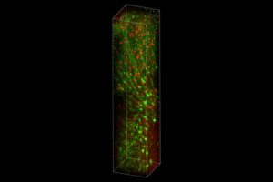
The team also developed a neuron-targeted excitation scheme that switches on the laser only as it scans identified cells. “The way we did this was through a series of very systematic, principled, and ground-up optimizations,” Vaziri said.
Together, these changes increased the fluorescence signal available for recording transient activity in individual neurons. The platform was tested in mice, and the paper reports recordings over fields of view approximately 230 by 230 µm2 at 8.6 Hz, reaching approximately 2 mm into the mouse cortex and hippocampus.
The advance expands access to circuits that previously required removal of overlying tissue for optical imaging, potentially helping researchers investigate how neuronal populations across deep brain regions coordinate their activity. It does not, however, make every deep structure accessible.
“There are regions of the striatum that are inaccessible, and there are still a number of deep brain nuclei which are much deeper that still require invasive approaches,” Vaziri added.
For now, the work demonstrates how coordinated optimization can bring three-photon imaging closer to its fundamental depth limit while preserving the tissue above the regions being studied.